Emergency injunction and tribunal order was served to WHO and all Criminal Defendants listed below to immediately cease and desist a criminal conspiracy to commit war crimes, genocide, crimes against humanity and to arrest and incarcerate these criminal defendants.
All Constitutional law enforcements officers worldwide are being served starting with Interpol and constitutional law enforcements in everyone of the 194 member nations under Writs of Mandamus ordering them to carry out their duties to arrest and incarcerate these criminal codefendants for unlawful and Genocidal acts in connection with the WHO Pandemic preparedness response and changes to the international health regulations.
Tribunal Writs of Mandamus is defined as: “The lawful term writ of mandamus refers to an order by a court to a lesser government official to perform an act required by law, which he has refused or neglected to do.
This type of court order is a remedy that may be sought if a governmental agency, public authority, or corporation in service of the government, fails or refuses to do its public or statutory duty.” This has been ordered by the Tribunal Under Articles 6 (Genocide) and 7 (Crimes Against Humanity) of the International Criminal Code.
List of Criminal Defendants 2009-2023 Lockstep Vaccination Genocidal Pandemic Criminal Co-conspiracy, including and not limited to individually and collectively:
Bill and Melinda Gates individually and the Bill and Melinda Gates Foundation.
World Health Organization (WHO) and Officers, Directors, Employees, and Agents Tedros Adhanom Ghebreyesus WHO Director General, Anthony Stephen Fauci.
World Economic Forum, Klaus Schwab and Officers, Directors, Employees, and Agents. Michael Bloomberg, David Rockefeller Jr, Warren Buffett, George Soros, Ted Turner, Oprah Winfrey, Rockefeller Foundation, Global Business Network (GBN), Peter Schwartz, Chairman GBN. Convicted Vaccination Genocidal Pandemic and Neural Monitoring Governmental Executive Defendants: Xi Jin
Ping, General Secretary of the Communist Party. Vladimir Vladimirovich Putin, President of the Russian Federation. Donald J. Trump, 45th President of the United States of America. Joseph R. Biden 46th President of the United States of America. Benjamin Netanyahu, Prime Minister of Israel. Boris Johnson, Prime Minister of the UK. Matt Hancock, UK Secretary of State for Health. The Crown Corporation and any and all of its Subsidiaries including and not limited to Serco. Justin Trudeau, Prime Minister of Canada. Scott Morrison, Prime Minister of Australia. Jacinda Arden, Prime Minister of New Zealand. Stefan Löfven Prime Minister of Sweden. Minister of Heath Lena Hallengren. Narendra Modi, Prime Minister of India. Emmanuel Jean-Michel Frédéric Macron, President of France. Edouard Phillipe, Prime Minister of France. Angela Merkel, Chancellor of Germany. António Guterres, Secretary General of the United Nations Organization. European Union, and President of the Commission, Ursula von der Leyen. President of the Parliament, David Sassoli. President of the European Council, Charles Michel. Michael Ryan,
WHO CEO’s at GCHQ-UK, NSA-USA and Bilderberg Group, all CEOs, Monarchies, and Members. Convicted Defendant Pandemic Vaccine Programs: Global Alliance Vaccine Initiative [GAVI], The Vaccine Alliance, including and not limited to its Pandemic Vaccine Program. Pandemic Vaccine Program United States of America, Operation Warp Speed and successor Pandemic Vaccine Program. World Health Organization COVAX, Pandemic Vaccine Program. Pandemic Vaccine Program People’s Republic of China, National Institutes for Food and Drug Control. Russian Federation, Rospotrebnadzor. Convicted Genocidal Vaccination Entities: Pfizer, AstraZeneca, Moderna, Johnson and Johnson, The Pirbright Institute.
02/24/23 Judge Pascal Najadi: They will all be arrested as soon as possible, Military and law enforcement worldwide are preparing for this to happen. Godspeed.
COVID Origins Hearing Wrap Up: Facts, Science, Evidence Point to a Wuhan Lab Leak
WASHINGTON—The Select Subcommittee on the Coronavirus Pandemic held a hearing on “Investigating the Origins of COVID-19” to gather facts about the origination of the virus that has claimed nearly seven million lives globally. At the hearing, several of the witnesses pointed to how the science, facts, and evidence point to a lab leak in Wuhan.
Key Hearing Takeaways
Knowing the origin of COVID-19 is fundamental to helping predict and prevent future pandemics.
Select Subcommittee Chairman Brad Wenstrup opened the hearing by emphasizing how knowing the origin of the virus is essential to helping predict and prevent future pandemics, protecting health and national security, and preparing the United States for the future. He pledged that the Select Subcommittee will thoroughly, responsibly, and honestly investigate the origin of COVID-19.
Dr. Jamie Metzl, Ph.D., senior fellow at the Atlantic Council said in an opening statement, “If we do not get to the bottom of what went wrong with the COVID-19 pandemic, if we fail in our efforts to fearlessly understand all shortcomings and shore up the vulnerabilities this crisis has so clearly exposed, the victims of the next pandemic, our children and grandchildren, will ask us why we failed to protect when we knew what was at stake and had the chance.”
Mounting evidence continues to show that COVID-19 may have originated from a lab in Wuhan, China.
Dr. Robert Redfield, former director of the U.S. Centers for Disease Control and Prevention (CDC), testified how science indicates COVID-19 infections were likely the result of an accidental lab leak in Wuhan. His conclusion is based on the biology of the virus itself and unusual actions in and around Wuhan in 2019, including gain-of-function research at the Wuhan Institute of Virology (WIV).
Nicholas Wade—the former science and health editor at the New York Times, and former editor of Science and Nature—testified how Drs. Fauci and Collins used unverified data to dismiss the lab leak theory in favor of natural transmission.
Jamie Metzl testified how China’s government destroyed samples, hid records, imprisoned Chinese journalists, prevented Chinese scientists from saying or writing anything on pandemic origins without prior government approval, actively spread misinformation, and prevented an evidence-based investigation.
The mainstream media downplayed—and even denied—the scientific theory that COVID-19 emerged from the WIV.
Nicholas Wade testified about the campaign to discredit the lab leak theory. He pointed out that scientists kept in line with the natural origin camp led by Drs. Fauci and Collins because of their dependence on government grants and that the media failed to challenge the forced narrative.
All witnesses agreed that the possibility of COVID-19 originating from a lab is not a conspiracy theory.
Member Highlights
Subcommittee Chairman Dr. Wenstrup (R-Ohio.) asked witnesses whether it is critical to investigate the origin of COVID-19. All witnesses answered yes. Chairman Wenstrup also raised concern about gain-of-function research, which Dr. Redfield defined during the hearing as altering a pathogen to increase either transmissibility or pathogenicity.
Subcommittee Chairman Wenstrup: “In your expert opinion was the Wuhan Institute conducting gain-of-function research on a batch of coronaviruses?”
Dr. Redfield: “Absolutely.”
Rep. Nicole Malliotakis (R-N.Y.) noted that after raising concerns to experts and the World Health Organization that COVID-19 may have originated in a lab in Wuhan, China, and urging Dr. Fauci to investigate the origins of the pandemic, Dr. Redfield was excluded from calls related to the origins of the pandemic.
Rep. Malliotakis: “Why do you think you were excluded from those calls?”
Dr. Redfield: “It was told to me that they wanted a single narrative and that I obviously had a different point of view.”
Dr. Redfield added: “If you really want to be truthful, it’s antithetical to science. Science has debate, and they squashed any debate.”
Scientists, including Dr. Fauci, then drafted a paper arguing COVID-19’s proximal origins to animals at a wet market.
Rep. Malliotakis: “Do you think that this paper does hide the truth?”
Dr. Redfield: “I think it’s an inaccurate paper that basically was part of a narrative that they were creating.”
Rep. Malliotakis also warned that the National Institutes of Health (NIH) may have been funding gain-of-function research on coronaviruses at the WIV.
Rep. Malliotakis: “Is it likely that American tax dollars funded the gain-of-function research that created this virus?”
Dr. Redfield: I think it did, not only from NIH, but from the State Department, USAID and DOD.”
Rep. Miller-Meeks (R-Iowa), who has expertise publishing in peer-reviewed scientific journals, asked why the scientific community dangerously suppressed evidence that COVID-19 may have originated from a lab.
“There is, as you said Dr. Metzl, extraordinary circumstantial evidence that this came from a lab.
“I don’t know why the authors didn’t want to state this, they did not want to have the scientific conversation and dialogue, why they wanted to obfuscate and suppress the truth, or even have a debate about the origins of COVID-19.
“Was it for personal financial gain? Was it to hide U.S. financial interest into the Wuhan Institute of Virology indirectly? Was it to suppress the revelation that there was perhaps gain-of-function research that had been prohibited in the United States? Or were they concerned that a conspiracy would develop that it was bioterrorism?
“I would state that their suppression and obfuscation has led to the exact mistrust and conspiracy theories that they may have tried to avoid.”
Rep. Debbie Lesko (R-Colo.) and Dr. Redfield discussed unusual actions at the WIV in September 2019.
Rep. Lesko: “Do you believe we can have certainty that the virus did not come from the Wuhan lab and that U.S. funding was not used for coronavirus research?”
Dr. Redfield: “Absolutely we cannot do that. It’s now declassified now, but in September 2019, three things happened in that lab. One, they deleted the sequences. That is highly irregular—researchers don’t usually like to do that. Second, they commanded the command and control of the lab from civilian control to military control. Highly unusual. And the third thing they did, which I think is really telling, is they let a contractor re-do the ventilation system in that laboratory. There is strong evidence there was a significant event in that laboratory in September 2019.”
Oversight Committee Chairman James Comer (R-Ky.) warned that the media downplayed, discredited, and silenced voices of experts sounding the alarm that COVID-19 may have originated from a lab in Wuhan, China.
Chairman Comer: “Would you agree that the scientific establishment used the media to downplay the lab leak theory?”
Mr. Wade: “I think the media was used in this particular campaign to establish the natural origin theory.
“The scientific community is very afraid to speak up on political issues. I think the reason is that government grants are handed out through the system of peer-reviewed committees. You don’t want any single scientist on your peer-review committee to vote against, because you won’t get your grant – it’s so competitive. Therefore, scientists are very reluctant to say anything that’s politically divisive or turn other scientists off against them. This means that they cannot be relied upon in the way that we would like them to be independent and forthright and call it as they see it.”
Comer: “Was there science available to make such an unequivocal statement against the possibility of a lab leak that early on in February of 2020?”
Witnesses Dr. Metzl, Mr. Wade, and Dr. Redfield all answered, “No.”
Comer: “Is the possibility COVID-19 leaked from a lab a conspiracy theory?”
Almamlouk R, Kashour T, Obeidat S, Bois MC, Maleszewski JJ, Omrani OA, Tleyjeh R, Berbari E, Chakhachiro Z, Zein-Sabatto B, Gerberi D, Tleyjeh IM; Cardiac Autopsy in COVID-19 Study Group; Paniz Mondolfi AE, Finn AV, Duarte-Neto AN, Rapkiewicz AV, Frustaci A, Keresztesi AA, Hanley B, Märkl B, Lardi C, Bryce C, Lindner D, Aguiar D, Westermann D, Stroberg E, Duval EJ, Youd E, Bulfamante GP, Salmon I, Auer J, Maleszewski JJ, Hirschbühl K, Absil L, Barton LM, Ferraz da Silva LF, Moore L, Dolhnikoff M, Lammens M, Bois MC, Osborn M, Remmelink M, Nascimento Saldiva PH, Jorens PG, Craver R, Aparecida de Almeida Monteiro R, Scendoni R, Mukhopadhyay S, Suzuki T, Mauad T, Fracasso T, Grimes Z. COVID-19-Associated cardiac pathology at the postmortem evaluation: a collaborative systematic review. Clin Microbiol Infect. 2022 Aug;28(8):1066-1075. doi: 10.1016/j.cmi.2022.03.021. Epub 2022 Mar 23. PMID: 35339672; PMCID: PMC8941843.
Abstract
Background
Many postmortem studies address the cardiovascular effects of COVID-19 and provide valuable information, but are limited by their small sample size.
Objectives
The aim of this systematic review is to better understand the various aspects of the cardiovascular complications of COVID-19 by pooling data from a large number of autopsy studies.
Data sources
We searched the online databases Ovid EBM Reviews, Ovid Embase, Ovid Medline, Scopus, and Web of Science for concepts of autopsy or histopathology combined with COVID-19, published between database inception and February 2021. We also searched for unpublished manuscripts using the medRxiv services operated by Cold Spring Harbor Laboratory.
Study eligibility criteria
Articles were considered eligible for inclusion if they reported human postmortem cardiovascular findings among individuals with a confirmed SARS coronavirus type 2 (CoV-2) infection.
Participants
Confirmed COVID-19 patients with post-mortem cardiovascular findings.
Interventions
None.
Methods
Studies were individually assessed for risk of selection, detection, and reporting biases. The median prevalence of different autopsy findings with associated interquartile ranges (IQRs).
Results
This review cohort contained 50 studies including 548 hearts. The median age of the deceased was 69 years. The most prevalent acute cardiovascular findings were myocardial necrosis (median: 100.0%; IQR, 20%–100%; number of studies = 9; number of patients = 64) and myocardial oedema (median: 55.5%; IQR, 19.5%–92.5%; number of studies = 4; number of patients = 46). The median reported prevalence of extensive, focal active, and multifocal myocarditis were all 0.0%. The most prevalent chronic changes were myocyte hypertrophy (median: 69.0%; IQR, 46.8%–92.1%) and fibrosis (median: 35.0%; IQR, 35.0%–90.5%). SARS-CoV-2 was detected in the myocardium with median prevalence of 60.8% (IQR 40.4-95.6%).
Conclusions
Our systematic review confirmed the high prevalence of acute and chronic cardiac pathologies in COVID-19 and SARS-CoV-2 cardiac tropism, as well as the low prevalence of myocarditis in COVID-19.
Preexisting cardiovascular comorbidities are prevalent among patients with COVID-19 and associated with a higher mortality rate [[1], [2], [3]]. For example, in the study reported by the Chinese Centre for Disease Control and Prevention describing the early experience with the epidemic in the Hubie province, patients with cardiovascular comorbidities had a case fatality rate of 10.5% compared with an overall cohort fatality rate of 2.3% [4]. There is also emerging robust evidence to suggest long-term cardiovascular sequalae after acute COVID-19 infection with an increased risk of incident conditions, including dysrhythmias, ischemic and nonischemic heart disease, myocarditis, and thromboembolic disease, among different COVID-19 disease severity groups compared with patients not infected with COVID-19 [5].
In addition, echocardiographic studies in populations infected with COVID-19 have demonstrated a high prevalence of ventricular dysfunction. In a prospective international study of 1216 patients with COVID-19, overall left and right ventricular dysfunction were reported in 39% and 33%, respectively [6]. Even in patients without preexisting cardiac disease, abnormal echocardiographic findings were evident in 46% of patients, with 13% manifesting severe abnormalities [6]. Acute myocardial injury manifesting as an elevation in cardiac troponins has been reported in 7% to 28% of patients with COVID-19 [[7], [8], [9], [10]]. Such acute cardiac injury was associated with higher overall mortality [10]. In a meta-analysis of 13 studies, the risk of death was high among patients with COVID-19 who had acute myocardial injury as defined by elevated serum troponins (risk ratio: 7.95; CI, 5.12–12.34; p <0.001; I2 = 65%) [11].
Several mechanisms have been proposed to explain acute myocardial injury and ventricular dysfunction in patients with COVID-19, including supply–demand mismatch secondary to hypoxemia and elevated cardiac demand, direct damage inflicted by inflammatory cytokines, microvascular dysfunction, myocarditis, coagulation abnormalities, and coronary artery plaque instability [12,13]. Other proposed mechanisms, such as vasospasm, microvascular thrombosis, and myocarditis, could be responsible for the ST-segment elevation [14].
A direct pathologic cardiovascular examination of decedents provides important information about the true frequency of cardiac complications among patients with COVID-19, and sheds light on possible pathologic mechanisms. Early on, small postmortem studies described evidence of myocardial inflammation associated with myocyte necrosis in patients with COVD-19 [15,16], as well as a possible direct SARS coronavirus type 2 (CoV-2) infection of the heart [17]. Moreover, nonspecific longstanding findings, such as cardiac hypertrophy and fibrosis, suggest underlying cardiovascular disease in a subset of these patients. Multiple subsequent studies have been published with varying sample sizes, methodologies, and findings. These studies provide valuable information about the nature of cardiac involvement in patients with COVID-19. However, their small sample sizes make deriving a clear picture of the true frequencies of cardiovascular complications in this novel disease challenging. In this international collaboration, we undertook a systematic review to better understand the pathologic cardiac findings in patients with COVID-19 at the time of postmortem evaluation.
We followed the Preferred Reporting Items for Systematic Reviews and Meta-analyses guidelines. The protocol of this review was registered in PROSPERO (CRD42020223551).
Literature search and study selection
The literature was searched by a medical librarian for the concepts of autopsy or histopathology combined with COVID-19. The search strategies were created using keywords and standardized index terms (Doc. S1). Searches were run in February 2021 in Ovid EBM Reviews, Ovid Embase (1974+), Ovid Medline (1946+, including ePUB ahead of print, in-process, and other nonindexed citations), Scopus (1970+), and Web of Science (1975+). We also searched for unpublished manuscripts using the medRxiv services operated by Cold Spring Harbor Laboratory. In addition, we searched Google Scholar and the references of eligible studies and review articles.
Articles were considered eligible for inclusion if they were studies with human participants and reported cardiac autopsy findings among individuals with a SARS-CoV-2 infection. We included studies published in any language.
Identification of studies
Two reviewers (RA and SO) examined the titles and abstracts of articles using the studies selection criteria. Then, they examined the full texts to confirm that each article met the eligibility criteria.
Data collection
Data were extracted by two reviewers (R.A. and S.O.) and in duplicates into a prespecified data collection form. Disagreements were discussed with the senior reviewers (I.T. and T.K.). Data were collected on the following prespecified outcomes: 1) Study location, study type, number of cases, patient selection, selection bias, and autopsy type; 2) baseline characteristics, including age, sex, ethnicity, body mass index, cause of death, days to death, and presence of comorbidities; 3) laboratory test values, including maximum serum troponin levels, serum brain natriuretic peptide, serum ferritin, and D-dimer levels; 4) cardiac autopsy findings; and 5) ultrastructural studies, including immunohistochemistry and electron microscopy. The Cardiac Autopsy in COVID-19 Study Group collaborators completed a data collection form (Doc. S2).
One author assessed the studies for risk of selection, detection, and reporting biases. Specifically, studies were evaluated on whether consecutively deceased patients with COVID-19 underwent a cardiac autopsy to reduce selection bias.
Statistical analyses
The number and percentage of patients manifesting different findings during cardiac autopsies were extracted from each study and confirmed with the studies’ authors. We initially planned to perform meta-analyses to obtain pooled estimates of the different findings’ prevalences. However, this was not possible due to the limited number of studies that performed consecutive cardiac autopsies. We report the median prevalence of cardiac autopsy findings across studies with sample sizes ≥5, with associated interquartile ranges (IQRs) (see Table 1 ).
Table 1
Summary of median prevalence of cardiac autopsy findings of studies with ≥5 patients
The search yielded 4760 results. We examined the entire text of 58 manuscripts after removing duplicates and screening the titles. However, eight studies were excluded, leaving 50 studies with 548 hearts in the final cohort (Fig. 1 ). Most studies were case reports (n = 13) or case series (n = 37; Doc. S3). Autopsy cases were acquired from manuscripts spanning experiences from 15 countries. The number of cases per study ranged from 1 to 80 (median: 4.5), and five cases were identified as reporting consecutive autopsies (encompassing 155 subjects) [[18], [19], [20], [21], [22]]. There were 42 minimally invasive autopsies, 102 partial autopsies, and 301 complete autopsies among the autopsies where completeness was stated or could be inferred.
Preferred Reporting Items for Systematic Reviews and Meta-analyses 2009 flow diagram.
Patient demographics, comorbidities, and cause of death
The median age of the deceased was 69 years (range, 22–97 years; n = 548), and 62% of cases were men (n = 338 of 548). The most common comorbidities were systemic hypertension (n = 298; 56%) and coronary artery disease (n = 252; 49%). Other less common comorbidities included chronic obstructive pulmonary disease, diabetes, obesity, chronic kidney disease, old myocardial infarction, dementia, malignancy, and sleep apnoea (Fig. 2 ). Elevated troponin was demonstrated in 55% of cases.
Bar chart showing reported comorbidities of deceased patients included in this cohort. CAD, coronary artery disease; CKD, chronic kidney disease; COPD, chronic obstructive pulmonary disease; MI, myocardial infarction. Data labels show the prevalence of reported comorbidities (can overlap in a single patient).
The cause of death was reported in 479 cases, with the most reported being respiratory in origin. However, in 62 cases, cardiac involvement was identified as a key factor in mortality. The median time from the onset of symptoms to death was 9 days (range, 0–71 days; n = 401).
Cardiovascular autopsy findings
General findings
Cardiac abnormalities were found in gross pathology or histology test results in almost all cases. Heart weights were available for 276 hearts (51%), with a median weight of 465 g (range, 238–1070 g).
SARS coronavirus type to infection of the heart
Nineteen studies [[17], [18], [19], [23], [24], [25], [26], [27], [28]] with 217 cases explored the presence and localization of SARS-CoV-2 infection in the heart using different modalities, including RT-PCR, immunohistochemistry, in situ hybridization, and electron microscopy. Ten studies [18,19,[26], [27], [28],30,32,33,35,37] with a total of 116 cases detected SARS-CoV-2 infection in the cardiac tissues in 70 cases with a median of 60.8% (IQR, 40.4%–95.6%; Fig. 3 ).
Box-and-whisker plot of cardiac autopsy findings of studies with ≥5 patients as median percentage prevalence and associated interquartile ranges. CAD, coronary artery disease.
Active replication of SARS-CoV-2 within the heart was determined using the RNA scope in situ hybridization technique looking for the presence of the negative strand of the SARS-CoV-2 viral RNA or through the identification of subgenomic RNA, both of which indicate active viral replication. Four investigators employed these techniques in 55 cases [18,27,28,33], and verified the presence of active SARS-CoV-2 viral replication in 15 hearts (27%).
Localization of SARS-CoV-2 within different cardiac cell compartments was studied by nine investigators [23,25,26,30,33,34,[36], [37], [38]] in 56 hearts from total of 95 cases using electron microscopy or immunohistochemistry. The presence of SARS-CoV-2 infection within the cardiomyocytes was reported in 11 hearts by four investigators [30,33,34,36]. SARS-CoV-2 infection was also detected in cardiac vascular endothelial cells in seven hearts and in cardiac fibroblasts in one heart [26,34]. On the other hand, other investigators [23,25,26,37,38] could not detect SARS-CoV-2 infection within any cell type in the heart.
Myocarditis
The majority of studies did not specify what definition of myocarditis was used. However, we inferred from the description of the histopathological findings that the Dallas criteria were used by most studies. Several investigators used immunohistochemical studies with different antibodies to identify subtypes of cellular infiltrates, but most did not use immunohistochemical criteria to diagnose myocarditis. In total, 36 cases had myocarditis and 16 had inflammatory infiltrates but no myocyte damage (Fig. 3).
Few cases reported extensive myocarditis, ranging from 0.0% to 19.3%, with a median of 0.0% across 10 studies [20,23,26,28,32,37,[39], [40], [41], [42]] with a total of 175 cases (Fig. 3). Grosse et al., who authored the only consecutive study to report a prevalence for this finding, did not find any cases of extensive myocarditis across 14 cases. Focal active myocarditis was reported by 13 studies [20,21,23,25,26,28,32,[37], [38], [39], [40],42,43], ranging from 0.0% to 55.5%. Nine studies [19,20,26,28,32,37,39,42,44] with total of 131 cases described multifocal myocarditis with a median prevalence of 0.0% (IQR, 0.0%–2.1%). Finally, 15 studies [18,[20], [21], [22],[26], [27], [28],32,35,37,39,40,42,44,45] with 279 cases reported infiltrates without myocyte damage with a median prevalence of 0.6% (range, 0.0%–28.9%; Fig. 3).
Other acute cardiac pathologic changes
Necrosis had the highest median reported prevalence across nine studies, of which none were considered consecutive studies, including 64 autopsies [28,29,31,34,40,43,[46], [47], [48]] with a median of 100% (n = 64; IQR, 20.0%–100%). This was followed by cardiac interstitial oedema (n = 46; median: 55%; IQR, 19.5%–92.5%) [22,27,30,42], with Duarte-Neto et al. reporting a prevalence of 90% across ten consecutive autopsies (Fig. 3).
Microvessel thrombi had the highest reported median prevalence across the category of thromboembolic disease among eight studies, reporting a similar prevalence across the studies [22,23,27,28,30,38,44,49] with 43 of 103 cases (median: 36.2%; IQR, 17.5%–61.7%). Alternatively, acute myocardial infarction had the lowest reported median prevalence in this category (median: 11.8%; IQR, 7.9%–13.8%; Fig. 3). Acute epi-pericarditis was reported with a median prevalence of 15.5% (IQR, 11.9%–19.2%) across six studies [28,30,33,38,39,41] in 29 of 110 cases, and small vessel vasculitis had a median reported prevalence of 28.6% (IQR, 16.0%–32.5%) across three studies [38,41,43] in 12 of 86 cases (Fig. 3). Other less frequently reported findings include single-cell ischemia in one of seven patients [35], myocyte ischemic degeneration with pyknosis in one case report [50], and contraction bands in one of three cases [51].
Chronic cardiac findings
Hypertrophy was the most common pathological finding with a median of 69.0% (IQR, 46.8%–92.1%) across 18 studies [[18], [19], [20],22,23,25,28,[31], [32], [33],35,38,39,[41], [42], [43],45,49] in 197 of 303 cases. Fibrosis was reported in 13 studies [20,22,25,27,28,[30], [31], [32],37,[42], [43], [44], [45]] with a median of 42.9% (IQR, 35.0%–90.5%) in 104 of 183 cases. Among these 13 studies, ten studies reported various details about the nature of fibrotic changes [20,25,27,28,30,31,37,42,44,45]. Two studies [42,44] reported on the severity of fibrosis with 32 cases (20 with mild and 8 with moderate fibrosis). Six studies [20,27,28,30,31,44] reported on the extent of fibrosis in a total of 98 cases. There was fibrosis in 46 of these cases, which was diffuse in 18 cases and focal or patchy in 28 cases. Replacement fibrosis was described by two authors [25,27] in eight cases. Eight studies [18,20,23,25,28,31,35,45,49] with 131 patients observed amyloidosis in 21 cases with a median prevalence of 13.6% (IQR, 9.8%–17.4%). The type of amyloidosis was reported in 14 of these cases, and determined to be transthyretin in 13 cases and amyloid P in one case, with patient age ranging between 71 and 96 years [20,23,28,31,35,45]. Other less-reported pathologies were chronic pericarditis, reported by three studies in 17 of 47 cases [28,33,43], and ischemic heart disease in 1 of 3 cases in one study [50].
This systematic review of pathology-derived cardiac changes in patients with COVID-19 included 50 studies with more than 500 cases, and was the effort of an international collaboration. The most prevalent chronic changes were myocardial hypertrophy, underlying coronary artery disease, and fibrosis (median: 69.0%, 46.2%, and 42.9%, respectively). The high prevalence of chronic cardiac pathologies among patients who died due to COVID-19 supports the findings from previously published epidemiologic studies [[1], [2], [3]].
Interestingly, another underlying cardiac disease, amyloidosis, was reported in a median of 13.6% of patients with COVID-19, with patient age ranging between 71 and 96 years. The overall prevalence of cardiac amyloidosis in an unselected, sequential autopsy population was reported at approximately 4% [23]. Conversely, a Finnish autopsy study of individuals age >85 years detected cardiac amyloidosis in 25% of cases [52]. Although cardiac amyloidosis prevalence almost certainly increases with patient age, cardiac amyloidosis is also likely underdiagnosed, particularly among patients with heart failure and preserved ejection fraction [[53], [54], [55]]. Nevertheless, the relatively high proportion of cardiac amyloidosis among decedents with COVID-19 compared with unselected autopsy rates suggests that this condition may render patients vulnerable to adverse outcomes from SARS-CoV-2 infection. This is further supported by the average age of patients with cardiac amyloidosis who died of COVID-19. Possible mechanisms for this complication have been proposed; however, decreased cardiac reserve innate to underlying cardiovascular disease, including amyloidosis, likely plays a significant role [56].
The prevalence of acute thromboembolic pathologies in descending frequency included microvessel thrombi (36.2%), pulmonary embolism (22.2%), cardiac large vessel thrombosis (14.3%), and acute myocardial infarction (11.8%). The increased cardiac and pulmonary vascular thrombi correlate strongly with the clinical evidence of increased thromboembolic phenomena in patients with COVID-19. Moreover, these thrombotic changes, along with the observed high prevalence of acute cardiac injuries (e.g. necrosis, oedema, and epi-pericarditis) are concordant with the clinically documented ventricular dysfunction [6] and serologic markers of cardiac injury, such as increased troponins [[7], [8], [9], [10]]. In fact, microvascular thrombosis has been cited as the causative agent of cardiac injury in most decedents with COVID-19 [57]. This finding dovetails with the lower prevalence of large vessel cardiac thrombosis, and is consistent with the coronary angiographic findings in patients with COVID-19, wherein a culprit lesion was not identified in more than 40% of patients with suspected acute myocardial infarction [14].
SARS-CoV-2 cardiac tropism
SARS-CoV-2 gains entry into the host cells through the binding of its spike protein to the angiotensin-converting enzyme 2 with the help of the host transmembrane protease serine 2 [58]. Both proteins have been shown to be expressed in the heart [23,[59], [60], [61]]. The predecessor of SARS-CoV-2 (SARS-CoV and its associated syndrome SARS) also uses the angiotensin-converting enzyme 2 protein for cell entry, and has been shown to infect the heart and induce inflammatory changes based on data from the first decade of the 21st century [61]. These findings, along with the clinical observations of acute cardiac injury among patients with COVID-19, prompted several investigators to address three important questions: 1) Is SARS-CoV-2 present in the hearts of decedents with COVID-19; 2) if so, which cell type(s) does SARS-CoV-2 infect; and 3) can SARS-CoV-2 replicate in heart tissues?
Twenty studies explored the presence of SARS-CoV-2 within the heart, the majority of which targeted the identification of SARS-CoV-2 RNA in heart tissues using RT-PCR or in situ hybridization. Other employed techniques included immunohistochemistry to identify SARS-CoV-2 proteins (e.g. spike or nucleocapsid protein), as well as electron microscopy. These investigators identified the presence of SARS-CoV-2 in almost 60% of the examined hearts. Additionally, a few investigators identified SARS-CoV-2 replication within heart tissues in several cases [18,27,28,33]. Furthermore, studies investigated cell-type localization of SARS-CoV-2 within the heart, and provided evidence of the presence of SARS-CoV-2 viral particles within the cardiomyocytes [30,33,34,36]. Bulfamante et al. observed degenerative changes in cardiomyocytes containing SARS-CoV-2 viral particles [33]. These findings are supported by a study that demonstrated SARS-CoV-2 infection and propagation in induced pluripotent stem cell-derived cardiomyocytes [62]. SARS-CoV-2 has also been found in vascular endothelial cells and cardiac fibroblasts [26,34]. These reports establish SARS-CoV-2 cardiac tropism, and present a possible link between SARS-CoV-2 and certain acute cardiac pathologies (e.g. myocarditis). However, although RT-PCR represents a time-efficient method to determine tissue positive for SARS-CoV-2, RT-PCR does not allow for tissue localization. Wong et al. suggested and attempted to validate a Fluorescence In Situ Hybridization (FISH) method using positive and negative controls by detecting endogenous human genes (POLR2A and PPIB) and a bacterial gene (dap gene of Bacillus subtilis) to allow for a tissue-specific analysis [63].
SARS-CoV-2-induced myocarditis
In this review, we subdivided the reported cardiac inflammatory processes in patients with COVID-19 into four categories based on the degree of myocardial involvement and the presence of associated myocyte damage. Overall, the prevalence of each category was low, with vast differences between individual studies that cannot be explained solely by the methodological differences of the studies, and likely indicate significant selection and reporting bias. The median reported prevalence of extensive myocarditis, multifocal active myocarditis, and focal active myocarditis were all 0.0%, and the median prevalence of inflammatory infiltrate without myocyte damage was 0.6%.
Regrettably, clinical correlation or pooled prevalence estimates in the included reported autopsy series were not possible due to the heterogenous results and paucity of clinical and imaging data provided. Nonetheless, reports of clinically diagnosed myocarditis with pathologic correlation have been reported among inpatients with COVID-19 [36,48,64]. Intriguingly, Gauchotte et al. demonstrated pathologic evidence of myocarditis without lung involvement, and further showed the presence of the SARS-CoV-2 genome in cardiomyocytes in this case [36]. This finding is concordant with other studies suggesting a greater degree of inflammation with viral presence in the heart [65].
The diagnosis of most cases of myocarditis included in this review were based on the Dallas criteria. This methodology, although widely accepted, is not without its inherent limitations. First and foremost, the Dallas criteria were developed to diagnose myocarditis by endomyocardial biopsy (EMB), not autopsy, wherein a more abundant amount of tissue is available for histologic evaluation. The generalization (and clinical significance) of small foci of myocyte damage within autopsy-derived cardiac tissue is challenging to ascertain. Other limitations of the Dallas criteria include significant interobserver variability and sampling errors [66,67].
Although less of an issue in autopsy-derived tissue, the focal nature of the disease leads to sampling errors that have been shown to compromise the sensitivity of the histopathological diagnosis of myocarditis by EMB [68,69]. Chow et al. had estimated that a mean of 17 samples per patient would be required to establish a diagnosis of myocarditis [69], which likely explains why examining an increased number of cardiac tissue blocks at the time of autopsy resulted in a greater likelihood of identifying focal myocarditis.
The overall low prevalence of myocarditis in patients with COVID-19 is of interest, particularly when placed in the greater context of the available literature. In a recent meta-analysis on the diagnosis of myocarditis by EMB (including 61 studies with 10,491 patients), the prevalence of myocarditis according to the Dallas criteria was 8.04% [70]. This diagnosis was made on the relatively limited amount of tissue provided by EMB. In contrast, this review shows a myocarditis prevalence of 8% in abundant available tissue, often comprising multiple blocks of myocardium with greater orders of magnitude in the amount of tissue to examine. The pretest factors among these data points differ, but underscores the overall low prevalence of myocarditis in COVID-19 deaths and is concordant with previous literature reviews on the topic [71].
Prior studies have shown the added sensitivity of immunohistochemistry in the diagnosis of myocarditis. Katzmann et al. showed that the sensitivity of the Dallas criteria in detecting myocarditis was much lower than when immunohistochemistry is utilized, with a detection rate of 50.8% (vs. 8.04% without immunohistochemistry) [70]. In another study of 84 cases of myocarditis based on the immunohistochemistry criteria, applying the Dallas criteria without immunohistochemistry would have categorized only 8% of these case as active myocarditis [72].
The true prevalence of myocarditis in COVID-19 remains very hard to determine from the current autopsy and imaging studies, the latter of which shows a discordantly high prevalence of myocarditis compared with postmortem examinations. A recent systematic review of cardiovascular magnetic resonance findings in COVID-19 including 199 patients showed that myocarditis was the most prevalent diagnosis (40.2%) [73]. Future studies should integrate clinical imaging and more rigorous and systematic autopsy studies to help resolve this issue. Such initiative should be conducted in the form of an international registry that uses a unified autopsy examination and imaging protocols in accordance with published guidelines [74,75].
Limitations
This systematic review included the largest number of studies and cases published to date on cardiac changes in fatal COVID-19 with both qualitative and quantitative analyses of different cardiac pathologies observed in COVID-19. However, our study has several limitations. First, the majority of the included studies were small. Second, these studies were heterogeneous in their methodologies and patient cultural origins, with very few studies performing consecutive autopsies, which makes meta-analyses unfeasible. Moreover, selection and reporting bias likely affected most included studies, as evidenced by the nonconsecutive nature of case recruitment and the very high differences between the studies in the perveances of reported pathologies.
Our systematic review confirmed the high prevalence of acute and chronic cardiac pathologies in the autopsy-derived hearts of decedents with COVID-19. These findings help explain observations from clinical epidemiologic studies, such as thromboembolic phenomena and acute myocardial injury. Our study also provides evidence for SARS-CoV-2 cardiac tropism, and confirmed the low prevalence of myocarditis in patients with COVID-19.
IMT designed the study. IMT, RA, SO, and MCB coordinated the study. DG designed and ran the literature search. RA, SO, OAO, RT, ZC, BZS, EB, and TK acquired the data, screened records, and extracted the data. IMT and OAO conducted the formal analyses. TK wrote the report with input from MCB and JJM. All authors provided critical conceptual input, analyzed and interpreted data, and critically revised the report.
Elie Berbari reports royalties or licenses from UTD of <$5000 per year. Amy V. Rapkiewicz reports payment for expert testimony by Eric Hack, Esq. Bruno Märkl reports grants or contracts from the German Registry of COVID-19 Autopsies, funded by the Federal Ministry of Health and the Federal Ministry of Education and Research within the framework of the network of university medicine. Diana Lindner reports support for the present manuscript, grants, and contracts from the German Centre for Cardiovascular Research, Deutsche Herzstiftung. Dirk Westermann reports consulting fees and payment or honoraria for lectures, presentations, speakers bureaus, manuscript writing, or educational events from Abiomed, Bayer, AstraZeneca, Novartis, and Medtronic. Klaus Hirschbühl reports grants or contracts from the German Registry of COVID-19 Autopsies, funded by the Federal Ministry of Health and the Federal Ministry of Education and Research within the framework of the network of university medicine. Luiz Fernando Ferraz da Silva reports grants or contracts, paid to their institution, from the Bill and Melinda Gates Foundation. Martin Lammens reports support for the present manuscript from the Belgian Fund for Scientific Research–Flanders. Michael Osborn reports grants or contracts from the North West London pathology research grant (£10,000), paid by their own institution, to set up a tissue bank and fund the procurement and use of tissue included in the current research, as well as payment or honoraria for lectures, presentations, speakers bureaus, manuscript writing, or educational events from Yale University ($300 for talking about COVID). He also reports a leadership or fiduciary role on a board, society, committee, or advocacy group as president of the Royal College of Pathologists, secretary of the BDIAP, president AAPT (all unpaid). Paulo Hilario Nascimento Saldiva reports support for the present manuscript from the Bill and Melinda Gates Foundation, Conselho Nacional de Desenvolvimento Científico e Tecnológico, Fundação de Amparo à Pesquisa do Estado de São Paulo, and Hospital das Clinicas da Faculdade de Medicina da Universidade de Paulo–HC Convida. Tadaka Suzuki reports grants or contracts from the Japan Agency for Medical Research and Development and Japan Society for the Promotion of Science (grants in aid). There was no funding source for this study.
The Cardiac Autopsy in COVID-19 Study Group consists of Alberto E. Paniz Mondolfi (Department of Pathology, Molecular and Cell-Based Medicine, New York, New York), Aloke V. Finn (CVPath Institute, Inc., Gaithersburg, Maryland; and University of Maryland, Baltimore, Maryland), Amaro Nunes Duarte-Neto (Departamento de Patologia, Faculdade de Medicina da Universidade de São Paulo, São Paulo, Brazil), Amy V. Rapkiewicz (NYU Winthrop Hospital, Department of Pathology, Long Island School of Medicine, Long Island, New York), Andrea Frustaci (Department of Clinical, Internal, Anesthesiologist and Cardiovascular Sciences, La Sapienza University, Rome, Italy; and Cellular and Molecular Cardiology Lab, IRCCS L. Spallanzani, Rome, Italy), Arthur-Atilla Keresztesi (Fogolyan Kristof Emergency County Hospital, Covasna County Institution of Forensic Medicine, Covasna, Romania), Brian Hanley (Department of Cellular Pathology, Northwest London Pathology, Imperial College London NHS Trust, London, UK; and Centre for Inflammatory Disease, Imperial College London, London, UK), Bruno Märkl (Institute of Pathology and Molecular Diagnostics, University Medical Center Augsburg, Augsburg, Germany), Christelle Lardi (University Center of Legal Medicine, Geneva University Hospital, Geneva, Switzerland), Clare Bryce (Icahn School of Medicine at Mount Sinai, New York, New York), Diana Lindner (Department of Cardiology, University Heart and Vascular Centre, Hamburg, Germany; and DZHK–German Center for Cardiovascular Research, Partner site, Hamburg/Kiel/Lübeck, Germany), Diego Aguiar (University Center of Legal Medicine, Geneva University Hospital, Geneva, Switzerland), Dirk Westermann (Department of Cardiology, University Heart and Vascular Centre, Hamburg, Germany; and DZHK–German Center for Cardiovascular Research, Partner site, Hamburg/Kiel/Lübeck, Germany), Edana Stroberg (Office of the Chief Medical Examiner, Oklahoma City, Oklahoma), Eric J. Duval (Office of the Chief Medical Examiner, Oklahoma City, Oklahoma), Esther Youd (Forensic Medicine and Science, University of Glasgow, Glasgow, UK), Gaetano Pietro Bulfamante (Unità di Anatomia Patologica, Dipartimento di Scienze della Salute, Università degli Studi di Milano, Milan, Italy; and Struttura Complessa di Anatomia Patologica e Genetica Medica, ASST Santi Paolo e Carlo, Milan, Italy), Isabelle Salmon (Department of Pathology, Erasme Hospital, Université Libre de Bruxelles (ULB), Brussels, Belgium; Centre Universitaire inter Régional d’expertise en Anatomie Pathologique Hospitalière, Jumet, Belgium; and DIAPath, Center for Microscopy and Molecular Imaging, ULB, Gosselies, Belgium), Johann Auer (Department of Cardiology and Intensive Care, St. Josef Hospital Braunau, Austria; and Department of Cardiology and Intensive Care, Kepler University of Medicine Linz, Austria), Joseph J. Maleszewski (Department of Laboratory Medicine and Pathology, Mayo Clinic, Rochester, Minnesota; and Department of Cardiovascular Medicine, Mayo Clinic, Rochester, Minnesota), Klaus Hirschbühl (Department of Hematology and Clinical Oncology, University Medical Center Augsburg, Augsburg, Germany), Lara Absil (Department of Pathology, Erasme Hospital, ULB, Brussels, Belgium), Lisa M. Barton (Office of the Chief Medical Examiner, Oklahoma City, Oklahoma), Luiz Fernando Ferraz da Silva (Departamento de Patologia, Faculdade de Medicina da Universidade de São Paulo, São Paulo, Brazil; and Serviço de Verificação de Óbitos da Capital, Universidade de São Paulo, São Paulo, Brazil), Luiza Moore (Department of Histopathology, Cambridge University Hospitals NHS Foundation Trust, Cambridge, UK; and Cancer, Ageing and Somatic Mutation, Wellcome Sanger Institute, Cambridge, UK), Marisa Dolhnikoff (Departamento de Patologia, Faculdade de Medicina da Universidade de São Paulo, São Paulo, Brazil), Martin Lammens (Department of Pathology, Antwerp University Hospital, University of Antwerp, Edegem, Belgium), Melanie C. Bois (Department of Laboratory Medicine and Pathology, Mayo Clinic, Rochester, Minnesota), Michael Osborn (Department of Cellular Pathology, Northwest London Pathology, Imperial College London NHS Trust, London, UK; Death Investigation Committee, Royal College of Pathologists, London, UK; and Nightingale NHS Hospital, London, UK), Myriam Remmelink (Department of Pathology, Erasme Hospital, ULB, Brussels, Belgium), Paulo Hilario Nascimento Saldiva (Departamento de Patologia, Faculdade de Medicina da Universidade de São Paulo, São Paulo, Brazil), Philippe G. Jorens (Infla-Med Research Consortium of Excellence, University of Antwerp, Antwerp, Belgium; Department of Medicine and Health Sciences, Laboratory of Experimental Medicine and Pediatrics, University of Antwerp, Antwerp, Belgium; and Department of Intensive Care Medicine, Antwerp University Hospital, University of Antwerp, Edegem, Belgium), Randall Craver (Children’s Hospital of New Orleans, New Orleans, Louisiana; and Louisiana State University Health Sciences Center, New Orleans, Louisiana), Renata Aparecida de Almeida Monteiro (Departamento de Patologia, Faculdade de Medicina da Universidade de São Paulo, São Paulo, Brazil), Roberto Scendoni (Institute of Legal Medicine, Department of Law, University of Macerata, Macerata, Italy), Sanjay Mukhopadhyay (Department of Pathology, Cleveland Clinic, Cleveland, Ohio), Tadaki Suzuki (National Institute of Infectious Diseases, Tokyo, Japan), Thais Mauad (Departamento de Patologia, Faculdade de Medicina da Universidade de São Paulo, São Paulo, Brazil), Tony Fracasso (University Center of Legal Medicine, Geneva University Hospital, Geneva, Switzerland), and Zachary Grimes (Icahn School of Medicine at Mount Sinai, New York, New York).
1. Mehta N., Kalra A., Nowacki A.S., Anjewierden S., Han Z., Bhat P., et al. Association of use of angiotensin-converting enzyme inhibitors and angiotensin II receptor blockers with testing positive for coronavirus disease 2019 (COVID-19) JAMA Cardiol. 2020;5:1020–1026. [PMC free article] [PubMed] [Google Scholar]
2. Reynolds H.R., Adhikari S., Pulgarin C., Troxel A.B., Iturrate E., Johnson S.B., et al. Renin-angiotensin-aldosterone system inhibitors and risk of COVID-19. N Engl J Med. 2020;382:2441–2448. [PMC free article] [PubMed] [Google Scholar]
3. Mancia G., Rea F., Ludergnani M., Apolone G., Corrao G. Renin-angiotensin-aldosterone system blockers and the risk of COVID-19. N Engl J Med. 2020;382:2431–2440. [PMC free article] [PubMed] [Google Scholar]
4. The Novel Coronavirus Pneumonia Emergency Response Epidemiology Team The epidemiological characteristics of an outbreak of 2019 novel coronavirus diseases (COVID-19)—China, 2020. China CDC Weekly. 2020;2:113–122. [PMC free article] [PubMed] [Google Scholar]
5. Xie Y., Xu E., Bowe B., Al-Aly Z. Long-term cardiovascular outcomes of COVID-19. Nat Med. 2022;28:583–590. [PMC free article] [PubMed] [Google Scholar]
6. Dweck M.R., Bularga A., Hahn R.T., Bing R., Lee K.K., Chapman A.R., et al. Global evaluation of echocardiography in patients with COVID-19. Eur Heart J Cardiovasc Imaging. 2020;21:949–958. [PMC free article] [PubMed] [Google Scholar]
7. Wang D., Hu B., Hu C., Zhu F., Liu X., Zhang J., et al. Clinical characteristics of 138 hospitalized patients with 2019 novel coronavirus-infected pneumonia in Wuhan, China. JAMA. 2020;323:1061–1069. [PMC free article] [PubMed] [Google Scholar]
8. Guo T., Fan Y., Chen M., Wu X., Zhang L., He T., et al. Cardiovascular implications of fatal outcomes of patients with coronavirus disease 2019 (COVID-19) JAMA Cardiol. 2020;5:811–818. [PMC free article] [PubMed] [Google Scholar]
9. Zhou F., Yu T., Du R., Fan G., Liu Y., Liu Z., et al. Clinical course and risk factors for mortality of adult inpatients with COVID-19 in Wuhan, China: a retrospective cohort study. Lancet. 2020;395:1054–1062. [PMC free article] [PubMed] [Google Scholar]
10. Shi S., Qin M., Shen B., Cai Y., Liu T., Yang F., et al. Association of cardiac injury with mortality in hospitalized patients with COVID-19 in Wuhan, China. JAMA Cardiol. 2020;5:802–810. [PMC free article] [PubMed] [Google Scholar]
11. Santoso A., Pranata R., Wibowo A., Al-Farabi M.J., Huang I., Antariksa B. Cardiac injury is associated with mortality and critically ill pneumonia in COVID-19: a meta-analysis. Am J Emerg Med. 2021;44:352–357. [PMC free article] [PubMed] [Google Scholar]
12. Kang Y., Chen T., Mui D., Ferrari V., Jagasia D., Scherrer-Crosbie M., et al. Cardiovascular manifestations and treatment considerations in COVID-19. Heart. 2020;106:1132. [PMC free article] [PubMed] [Google Scholar]
13. Libby P. The heart in COVID-19: primary target or secondary bystander? JACC Basic Transl Sci. 2020;5:537–542. [PMC free article] [PubMed] [Google Scholar]
14. Stefanini G.G., Montorfano M., Trabattoni D., Andreini D., Ferrante G., Ancona M., et al. ST-elevation myocardial infarction in patients with COVID-19: clinical and angiographic outcomes. Circulation. 2020;141:2113–2116. [PMC free article] [PubMed] [Google Scholar]
15. Chen C., Zhou Y., Wang D.W. SARS-CoV-2: a potential novel etiology of fulminant myocarditis. Herz. 2020;45:230–232. [PMC free article] [PubMed] [Google Scholar]
16. Inciardi R.M., Lupi L., Zaccone G., Italia L., Raffo M., Tomasoni D., et al. Cardiac involvement in a patient with coronavirus disease 2019 (COVID-19) JAMA Cardiol. 2020;5:819–824. [PMC free article] [PubMed] [Google Scholar]
17. Tian S., Xiong Y., Liu H., Niu L., Guo J., Liao M., et al. Pathological study of the 2019 novel coronavirus disease (COVID-19) through postmortem core biopsies. Mod Pathol. 2020;33:1007–1014. [PMC free article] [PubMed] [Google Scholar]
18. Lindner D., Fitzek A., Bräuninger H., Aleshcheva G., Edler C., Meissner K., et al. Association of cardiac infection with SARS-CoV-2 in confirmed COVID-19 autopsy cases. JAMA Cardiol. 2020;5:1281–1285. [PMC free article] [PubMed] [Google Scholar]
19. Wichmann D., Sperhake J.-P., Lütgehetmann M., Steurer S., Edler C., Heinemann A., et al. Autopsy findings and venous thromboembolism in patients with COVID-19. Ann Intern Med. 2020;173:268–277. [PMC free article] [PubMed] [Google Scholar]
20. Grosse C., Grosse A., Salzer H.J.F., Dünser M.W., Motz R., Langer R. Analysis of cardiopulmonary findings in COVID-19 fatalities: high incidence of pulmonary artery thrombi and acute suppurative bronchopneumonia. Cardiovasc Pathol. 2020;49:107263. [PMC free article] [PubMed] [Google Scholar]
21. Edler C., Schröder A.S., Aepfelbacher M., Fitzek A., Heinemann A., Heinrich F., et al. Dying with SARS-CoV-2 infection-an autopsy study of the first consecutive 80 cases in Hamburg, Germany. Int J Leg Med. 2020;134:1275–1284. [PMC free article] [PubMed] [Google Scholar]
22. Duarte-Neto A.N., Monteiro R.A.A., da Silva L.F.F., Malheiros D., de Oliveira E.P., Theodoro-Filho J., et al. Pulmonary and systemic involvement in COVID-19 patients assessed with ultrasound-guided minimally invasive autopsy. Histopathology. 2020;77:186–197. [PMC free article] [PubMed] [Google Scholar]
23. Bois M.C., Boire N.A., Layman A.J., Aubry M.-C., Alexander M.P., Roden A.C., et al. COVID-19-associated nonocclusive fibrin microthrombi in the heart. Circulation. 2021;143:230–243. [PMC free article] [PubMed] [Google Scholar]
24. Bösmüller H., Traxler S., Bitzer M., Häberle H., Raiser W., Nann D., et al. The evolution of pulmonary pathology in fatal COVID-19 disease: an autopsy study with clinical correlation. Virchows Archiv. 2020;477:349–357. [PMC free article] [PubMed] [Google Scholar]
25. Bradley B.T., Maioli H., Johnston R., Chaudhry I., Fink S.L., Xu H., et al. Histopathology and ultrastructural findings of fatal COVID-19 infections in Washington State: a case series. Lancet. 2020;396:320–332. [PMC free article] [PubMed] [Google Scholar]
26. Fox S.E., Li G., Akmatbekov A., Harbert J.L., Lameira F.S., Brown J.Q., et al. Unexpected features of cardiac pathology in COVID-19 infection. Circulation. 2020;142:1123–1125. [PubMed] [Google Scholar]
27. Brook O.R., Piper K.G., Mercado N.B., Gebre M.S., Barouch D.H., Busman-Sahay K., et al. Feasibility and safety of ultrasound-guided minimally invasive autopsy in COVID-19 patients. Abdom Radiol (NY) 2021;46:1263–1271. [PMC free article] [PubMed] [Google Scholar]
28. Hanley B., Naresh K.N., Roufosse C., Nicholson A.G., Weir J., Cooke G.S., et al. Histopathological findings and viral tropism in UK patients with severe fatal COVID-19: a post-mortem study. Lancet Microbe. 2020;1:e245–e253. [PMC free article] [PubMed] [Google Scholar]
29. Jacobs W., Lammens M., Kerckhofs A., Voets E., Van San E., Van Coillie S., et al. Fatal lymphocytic cardiac damage in coronavirus disease 2019 (COVID-19): autopsy reveals a ferroptosis signature. ESC Heart Fail. 2020;7:3772–3781. [PMC free article] [PubMed] [Google Scholar]
30. Schurink B., Roos E., Radonic T., Barbe E., Bouman C.S.C., de Boer H.H., et al. Viral presence and immunopathology in patients with lethal COVID-19: a prospective autopsy cohort study. Lancet Microbe. 2020;1:e290–e299. [PMC free article] [PubMed] [Google Scholar]
31. Menter T., Haslbauer J.D., Nienhold R., Savic S., Hopfer H., Deigendesch N., et al. Postmortem examination of COVID-19 patients reveals diffuse alveolar damage with severe capillary congestion and variegated findings in lungs and other organs suggesting vascular dysfunction. Histopathology. 2020;77:198–209. [PMC free article] [PubMed] [Google Scholar]
32. Remmelink M., De Mendonça R., D’Haene N., De Clercq S., Verocq C., Lebrun L., et al. Unspecific post-mortem findings despite multiorgan viral spread in COVID-19 patients. Crit Care. 2020;24:495. [PMC free article] [PubMed] [Google Scholar]
33. Bulfamante G.P., Perrucci G.L., Falleni M., Sommariva E., Tosi D., Martinelli C., et al. Evidence of SARS-CoV-2 transcriptional activity in cardiomyocytes of COVID-19 patients without clinical signs of cardiac involvement. Biomedicines. 2020;8:626. [PMC free article] [PubMed] [Google Scholar]
34. Dolhnikoff M., Ferreira Ferranti J., de Almeida Monteiro R.A., Duarte-Neto A.N., Soares Gomes-Gouvêa M., Viu Degaspare N., et al. SARS-CoV-2 in cardiac tissue of a child with COVID-19-related multisystem inflammatory syndrome. Lancet Child Adolesc Health. 2020;4:790–794. [PMC free article] [PubMed] [Google Scholar]
35. Recalde-Zamacona B., García-Tobar L., Argueta A., Álvarez L., De Andrea C.E., Fernández Alonso M., et al. Histopathological findings in fatal COVID-19 severe acute respiratory syndrome: preliminary experience from a series of 10 Spanish patients. Thorax. 2020;75:1116–1118. [PMC free article] [PubMed] [Google Scholar]
36. Gauchotte G., Venard V., Segondy M., Cadoz C., Esposito-Fava A., Barraud D., et al. SARS-Cov-2 fulminant myocarditis: an autopsy and histopathological case study. Int J Leg Med. 2021;135:577–581. [PMC free article] [PubMed] [Google Scholar]
37. Martines R.B., Ritter J.M., Matkovic E., Gary J., Bollweg B.C., Bullock H., et al. Pathology and pathogenesis of SARS-CoV-2 associated with fatal coronavirus disease, United States. Emerg Infect Dis. 2020;26:2005–2015. [PMC free article] [PubMed] [Google Scholar]
38. Rapkiewicz A.V., Mai X., Carsons S.E., Pittaluga S., Kleiner D.E., Berger J.S., et al. Megakaryocytes and platelet-fibrin thrombi characterize multi-organ thrombosis at autopsy in COVID-19: a case series. EClinicalMedicine. 2020;24:100434. [PMC free article] [PubMed] [Google Scholar]
39. Schaller T., Hirschbühl K., Burkhardt K., Braun G., Trepel M., Märkl B., et al. Postmortem examination of patients with COVID-19. JAMA. 2020;323:2518–2520. [PMC free article] [PubMed] [Google Scholar]
40. Beigmohammadi M.T., Jahanbin B., Safaei M., Amoozadeh L., Khoshavi M., Mehrtash V., et al. Pathological findings of postmortem biopsies from lung, heart, and liver of 7 deceased COVID-19 patients. Int J Surg Pathol. 2021;29:135–145. [PMC free article] [PubMed] [Google Scholar]
41. del Nonno F., Frustaci A., Verardo R., Chimenti C., Nicastri E., Antinori A., et al. Virus-negative myopericarditis in human coronavirus infection. Circ Heart Fail. 2020;13 [PMC free article] [PubMed] [Google Scholar]
42. De Michele S., Sun Y., Yilmaz M.M., Katsyv I., Salvatore M., Dzierba A.L., et al. Forty postmortem examinations in COVID-19 patients. Am J Clin Pathol. 2020;154:748–760. [PMC free article] [PubMed] [Google Scholar]
43. Falasca L., Nardacci R., Colombo D., Lalle E., Di Caro A., Nicastri E., et al. Postmortem findings in Italian patients with COVID-19: a descriptive full autopsy study of cases with and without comorbidities. J Infect Dis. 2020;222:1807–1815. [PMC free article] [PubMed] [Google Scholar]
44. Basso C., Leone O., Rizzo S., De Gaspari M., van der Wal A.C., Aubry M.C., et al. Pathological features of COVID-19-associated myocardial injury: a multicentre cardiovascular pathology study. Eur Heart J. 2020;41:3827–3835. [PMC free article] [PubMed] [Google Scholar]
45. Lax S.F., Skok K., Zechner P., Kessler H.H., Kaufmann N., Koelblinger C., et al. Pulmonary arterial thrombosis in COVID-19 with fatal outcome: results from a prospective, single-center, clinicopathologic case series. Ann Intern Med. 2020;173:350–361. [PMC free article] [PubMed] [Google Scholar]
46. Guagliumi G., Sonzogni A., Pescetelli I., Pellegrini D., Finn A.V. Microthrombi and ST-segment–elevation myocardial infarction in COVID-19. Circulation. 2020;142:804–809. [PMC free article] [PubMed] [Google Scholar]
47. Buja L.M., Wolf D.A., Zhao B., Akkanti B., McDonald M., Lelenwa L., et al. The emerging spectrum of cardiopulmonary pathology of the coronavirus disease 2019 (COVID-19): report of 3 autopsies from Houston, Texas, and review of autopsy findings from other United States cities. Cardiovasc Pathol. 2020;48:107233. [PMC free article] [PubMed] [Google Scholar]
48. Craver R., Huber S., Sandomirsky M., McKenna D., Schieffelin J., Finger L. Fatal eosinophilic myocarditis in a healthy 17-year-old male with severe acute respiratory syndrome coronavirus 2 (SARS-CoV-2c) Fetal Pediatr Pathol. 2020;39:263–268. [PMC free article] [PubMed] [Google Scholar]
49. Keresztesi A.A., Perde F., Ghita-Nanu A., Radu C.C., Negrea M., Keresztesi G. Post-mortem diagnosis and autopsy findings in SARS-CoV-2 infection: Forensic case series. Diagnostics. 2020;10:1070. [PMC free article] [PubMed] [Google Scholar]
50. Wang X.X., Shao C., Huang X.J., Sun L., Meng L.J., Liu H., et al. Histopathological features of multiorgan percutaneous tissue core biopsy in patients with COVID-19. J Clin Pathol. 2021;74:522. [PMC free article] [PubMed] [Google Scholar]
51. Youd E., Moore L. COVID-19 autopsy in people who died in community settings: the first series. J Clin Pathol. 2020;73:840. [PubMed] [Google Scholar]
52. Tanskanen M., Peuralinna T., Polvikoski T., Notkola I.L., Sulkava R., Hardy J., et al. Senile systemic amyloidosis affects 25% of the very aged and associates with genetic variation in alpha2-macroglobulin and tau: a population-based autopsy study. Ann Med. 2008;40:232–239. [PubMed] [Google Scholar]
53. González-López E., Gallego-Delgado M., Guzzo-Merello G., de Haro-del Moral F.J., Cobo-Marcos M., Robles C., et al. Wild-type transthyretin amyloidosis as a cause of heart failure with preserved ejection fraction. Eur Heart J. 2015;36:2585–2594. [PubMed] [Google Scholar]
54. Bennani Smires Y., Victor G., Ribes D., Berry M., Cognet T., Méjean S., et al. Pilot study for left ventricular imaging phenotype of patients over 65 years old with heart failure and preserved ejection fraction: the high prevalence of amyloid cardiomyopathy. Int J Cardiovasc Imaging. 2016;32:1403–1413. [PubMed] [Google Scholar]
55. Mohammed S.F., Mirzoyev S.A., Edwards W.D., Dogan A., Grogan D.R., Dunlay S.M., et al. Left ventricular amyloid deposition in patients with heart failure and preserved ejection fraction. JACC Heart Fail. 2014;2:113–122. [PMC free article] [PubMed] [Google Scholar]
56. Brannagan T.H., 3rd, Auer-Grumbach M., Berk J.L., Briani C., Bril V., Coelho T., et al. ATTR amyloidosis during the COVID-19 pandemic: insights from a global medical roundtable. Orphanet J Rare Dis. 2021;16:204. [PMC free article] [PubMed] [Google Scholar]
57. Pellegrini D., Kawakami R., Guagliumi G., Sakamoto A., Kawai K., Gianatti A., et al. Microthrombi as a major cause of cardiac injury in COVID-19. Circulation. 2021;143:1031–1042. [PubMed] [Google Scholar]
58. Hoffmann M., Kleine-Weber H., Schroeder S., Krüger N., Herrler T., Erichsen S., et al. SARS-CoV-2 cell entry depends on ACE2 and TMPRSS2 and is blocked by a clinically proven protease inhibitor. Cell. 2020;181:271–280. e8. [PMC free article] [PubMed] [Google Scholar]
59. Liu H., Gai S., Wang X., Zeng J., Sun C., Zhao Y., et al. Single-cell analysis of SARS-CoV-2 receptor ACE2 and spike protein priming expression of proteases in the human heart. Cardiovasc Res. 2020;116:1733–1741. [PMC free article] [PubMed] [Google Scholar]
60. Dong M., Zhang J., Ma X., Tan J., Chen L., Liu S., et al. ACE2, TMPRSS2 distribution and extrapulmonary organ injury in patients with COVID-19. Biomed Pharmacother. 2020;131:110678. [PMC free article] [PubMed] [Google Scholar]
61. Oudit G.Y., Kassiri Z., Jiang C., Liu P.P., Poutanen S.M., Penninger J.M., et al. SARS-coronavirus modulation of myocardial ACE2 expression and inflammation in patients with SARS. Eur J Clin Invest. 2009;39:618–625. [PMC free article] [PubMed] [Google Scholar]
62. Pérez-Bermejo J.A., Kang S., Rockwood S.J., Simoneau C.R., Joy D.A., Ramadoss G.N., et al. SARS-CoV-2 infection of human iPSC-derived cardiac cells predicts novel cytopathic features in hearts of COVID-19 patients. bioRxiv. 2020 [Epub ahead of print] [PMC free article] [PubMed] [Google Scholar]
63. Wong D.W.L., Klinkhammer B.M., Djudjaj S., Villwock S., Timm M.C., Buhl E.M., et al. Multisystemic cellular tropism of SARS-CoV-2 in autopsies of COVID-19 patients. Cells. 2021;10:1900. [PMC free article] [PubMed] [Google Scholar]
64. Auer J., Neuhierl F., Hetzmann Z. COVID-19-related fatal myocarditis in a 42-year-old female patient. Cardiol J. 2020;27:642–643. [PMC free article] [PubMed] [Google Scholar]
65. Bearse M., Hung Y.P., Krauson A.J., Bonanno L., Boyraz B., Harris C.K., et al. Factors associated with myocardial SARS-CoV-2 infection, myocarditis, and cardiac inflammation in patients with COVID-19. Mod Pathol. 2021;34:1345–1357. [PMC free article] [PubMed] [Google Scholar]
66. Mason J.W., O’Connell J.B., Herskowitz A., Rose N.R., McManus B.M., Billingham M.E., et al. A clinical trial of immunosuppressive therapy for myocarditis. N Engl J Med. 1995;333:269–275. [PubMed] [Google Scholar]
67. Shanes J.G., Ghali J., Billingham M.E., Ferrans V.J., Fenoglio J.J., Edwards W.D., et al. Interobserver variability in the pathologic interpretation of endomyocardial biopsy results. Circulation. 1987;75:401–405. [PubMed] [Google Scholar]
68. Hauck A.J., Kearney D.L., Edwards W.D. Evaluation of postmortem endomyocardial biopsy specimens from 38 patients with lymphocytic myocarditis: implications for role of sampling error. Mayo Clin Proc. 1989;64:1235–1245. [PubMed] [Google Scholar]
69. Chow L.H., Radio S.J., Sears T.D., McManus B.M. Insensitivity of right ventricular endomyocardial biopsy in the diagnosis of myocarditis. J Am Coll Cardiol. 1989;14:915–920. [PubMed] [Google Scholar]
70. Katzmann J.L., Schlattmann P., Rigopoulos A.G., Noutsias E., Bigalke B., Pauschinger M., et al. Meta-analysis on the immunohistological detection of inflammatory cardiomyopathy in endomyocardial biopsies. Heart Fail Rev. 2020;25:277–294. [PubMed] [Google Scholar]
71. Halushka M.K., Vander Heide R.S. Myocarditis is rare in COVID-19 autopsies: cardiovascular findings across 277 postmortem examinations. Cardiovasc Pathol. 2021;50:107300. [PMC free article] [PubMed] [Google Scholar]
72. Wojnicz R., Nowalany-Kozielska E., Wojciechowska C., Glanowska G., Wilczewski P., Niklewski T., et al. Randomized, placebo-controlled study for immunosuppressive treatment of inflammatory dilated cardiomyopathy. Circulation. 2001;104:39–45. [PubMed] [Google Scholar]
73. Ojha V., Verma M., Pandey N.N., Mani A., Malhi A.S., Kumar S., et al. Cardiac magnetic resonance imaging in coronavirus disease 2019 (COVID-19): a systematic review of cardiac magnetic resonance imaging findings in 199 patients. J Thorac Imaging. 2021;36:73–83. [PubMed] [Google Scholar]
74. Basso C., Aguilera B., Banner J., Cohle S., d’Amati G., de Gouveia R.H., et al. Guidelines for autopsy investigation of sudden cardiac death: 2017 update from the Association for European Cardiovascular Pathology. Virchows Arch. 2017;471:691–705. [PMC free article] [PubMed] [Google Scholar]
75. Caforio A.L.P., Pankuweit S., Arbustini E., Basso C., Gimeno-Blanes J., Felix S.B., et al. Current state of knowledge on aetiology, diagnosis, management, and therapy of myocarditis: a position statement of the European Society of Cardiology Working Group on Myocardial and Pericardial Diseases. Eur Heart J. 2013;34:2636–2648. [PubMed] [Google Scholar]
Testimony of Meryl Nass, MD before the Health and Human Services Committee January 11, 2022 Honorable Chairpersons, Members and Senators, I write in support of LD 867. There are many reasons why preventing COVID vaccine mandates until adequate, sufficient safety studies have been performed is the right decision for this committee and legislature. 1. COVID vaccines are experimental Let me say, first, that no matter what claims have been made regarding these vaccines, they are not “safe and effective.” “Safe and effective” is an FDA ‘term of art’1 that may only be applied to licensed drugs and vaccines. All currently available COVID vaccines in the United States are unlicensed and experimental, a.k.a. investigational. Medicines and vaccines are either licensed products or experimental products. There is no gray area between them in US law. Whether or not research is explicitly conducted, the use of experimental products (including those issued under an Emergency Use Authorization) falls under the Nuremberg Code and under US law regulating experimental drugs. As former FDA Commissioner Stephen Hahn himself noted, “EUA products are still considered investigational.”
2According to 21CFR Subchapter D Part 312:3 “an experiment is any use of a drug except for the use of a marketed drug in the course of medical practice.” Vaccines are considered a subset of drugs by FDA.4 And the use of unlicensed, Emergency Use Authorized vaccines is thus, by definition, experimental. US law requires that humans receiving experimental products must provide written informed consent.5 However, when the PREP Act creating Emergency Use Authorizations (EUAs) was written, this requirement was loosened slightly for emergencies in which EUA products would be used. The required disclosures when using EUAs were specified below. Please note the option to accept or refuse. 21 U.S. Code § 360bbb–3 – Authorization for medical products for use in emergencies6 (ii) Appropriate conditions designed to ensure that individuals to whom the product is administered are informed— 1 https://www.fda.gov/science-research/risk-communication/fdas-risk-communication-research-agenda 2 https://www.usatoday.com/story/news/2020/11/24/fda-commissioner-stephen-hahn-timing-safety-covid-19- vaccine/6393865002/ 3https://www.accessdata.fda.gov/scripts/cdrh/cfdocs/cfcfr/cfrsearch.cfm?fr=312.3#:~:text=Clinical%20investigation %20means%20any%20experiment,the%20course%20of%20medical%20practice. 4 https://www.ncbi.nlm.nih.gov/pmc/articles/PMC7152379/ 5 https://www.ecfr.gov/on/2018-07-19/title-45/subtitle-A/subchapter-A/part-46#sp45.1.46.a 6 https://www.law.cornell.edu/uscode/text/21/360bbb-32 (I) that the Secretary has authorized the emergency use of the product; (II) of the significant known and potential benefits and risks of such use, and of the extent to which such benefits and risks are unknown; and (III) of the option to accept or refuse administration of the product, of the consequences, if any, of refusing administration of the product, and of the alternatives to the product that are available and of their benefits and risks. All Moderna, Janssen (Johnson and Johnson) and all childhood Pfizer-BioNTech vaccines are being used under EUAs. And while the adult Pfizer-BioNTech vaccine is supposed to be licensed with brand name Comirnaty, in fact the Pfizer vaccines being used in the US today are EUA products as well. 2. While FDA licensed Comirnaty, the only approved COVID vaccine, only Emergency Use Authorized (experimental) vaccines are being used Despite claims to the contrary, the only vaccine currently available in the US is the Pfizer- BioNTech, not the licensed and branded Comirnaty. The Pfizer-BioNTech vaccine is authorized under an Emergency Use Authorization, which provides a broad liability shield to the manufacturer, distributor, administrator, program planner, and virtually anyone else involved in the vaccination process. The branded product, on the other hand, is subject to ordinary liability claims at the present time. Exactly three weeks after FDA issued Comirnaty a license, the National Library of Medicine, part of the NIH, posted information that Pfizer was not planning to make Comirnaty available in the US while the EUA vaccine was still available:7 “SEPTEMBER 13, 2021 Pfizer received FDA BLA license for its COVID-19 vaccine Pfizer received FDA BLA license on 8/23/2021 for its COVID-19 vaccine for use in individuals 16 and older (COMIRNATY). At that time, the FDA published a BLA package insert that included the approved new COVID-19 vaccine tradename COMIRNATY and listed 2 new NDCs (0069-1000-03, 0069-1000-02) and images of labels with the new tradename. At present, Pfizer does not plan to produce any product with these new NDCs and labels over the next few months while EUA authorized product is still available and being made available for U.S. distribution. As such, the CDC, AMA, and drug compendia may not publish these new codes until Pfizer has determined when the product will be produced with the BLA labels.” FDA extended the vaccine’s EUA authorization on the same day it licensed the vaccine. 7 https://dailymed.nlm.nih.gov/dailymed/dailymed-announcements-details.cfm?date=2021-09-133 FDA appears to have been acceding to the White House demand that the vaccine be licensed, in order for it to be mandated for large sectors of the US population. Under an EUA, which specifies that potential recipients have the right to refuse,8 mandates cannot be imposed. So a license was issued, allowing the administration to inform the public that the vaccine was fully approved and licensed. But in fact, the public was unable to access the licensed vaccine. Why was this convoluted regulatory process performed? While under EUA, Pfizer has an almost bulletproof liability shield. According to the Congressional Research Service (CRS) on September 23, 2021,9 “courts have characterized PREP Act immunity as ‘sweeping.'” The CRS explains, “the PREP Act immunizes a covered person from legal liability for all claims for loss relation to the administration or use of a covered countermeasure.” 3.FDA instructed Pfizer-BioNTech that FDA’s Congressionally-mandated databases are inadequate to assess the danger of myocarditis (and other potential COVID vaccine side effects) and therefore Pfizer-BioNTech must perform studies to evaluate these risks over the next six years On the day FDA issued a license for Comirnaty, August 23, 2021, FDA instructed Pfizer- BioNTech that it did NOT have sufficient information on serious potential risks of the product, and required Pfizer and BioNTech, the manufacturers, to conduct a series of studies to assess these potential risks.10 These studies were to be performed on both products: the licensed Comirnaty and the EUA Pfizer-BioNTech vaccine. Note that they include the requirement for a safety study in pregnancy, which will not be completed until December 31, 2025. I have reproduced part of what FDA wrote about these required safety studies below, directly from pages 6-11 of the FDA approval letter sent to BioNTech, linked below. FDA’s admission that it cannot assess these safety risks, and that up to 6 years will be taken to study them, provides us with additional de facto evidence that the Pfizer vaccines cannot be termed safe, as many of the fundamental safety studies are only now getting started. https://www.fda.gov/media/151710/download “POSTMARKETING REQUIREMENTS UNDER SECTION 505(o) Section 505(o) of the Federal Food, Drug, and Cosmetic Act (FDCA) authorizes FDA to require holders of approved drug and biological product applications to conduct postmarketing studies and clinical trials for certain purposes, if FDA makes certain findings required by the statute (section 505(o)(3)(A), 21 U.S.C. 355(o)(3)(A)). We have determined that an analysis of spontaneous postmarketing adverse events reported under section 505(k)(1) of the FDCA will not be sufficient to assess known 8 https://www.law.cornell.edu/uscode/text/21/360bbb-3 9 https://crsreports.congress.gov/product/pdf/LSB/LSB10443 10 https://www.fda.gov/media/151710/download4 serious risks of myocarditis and pericarditis and identify an unexpected serious risk of subclinical myocarditis. Furthermore, the pharmacovigilance system that FDA is required to maintain under section 505(k)(3) of the FDCA is not sufficient to assess these serious risks. Therefore, based on appropriate scientific data, we have determined that you are required to conduct the following studies: 4. Study C4591009, entitled “A Non-Interventional Post-Approval Safety Study of the Pfizer-BioNTech COVID-19 mRNA Vaccine in the United States,” to evaluate the occurrence of myocarditis and pericarditis following administration of COMIRNATY. We acknowledge the timetable you submitted on August 21, 2021, which states that you will conduct this study according to the following schedule: Final Protocol Submission: August 31, 2021 Monitoring Report Submission: October 31, 2022 Interim Report Submission: October 31, 2023 Study Completion: June 30, 2025 Final Report Submission: October 31, 2025 5. Study C4591021, entitled “Post Conditional Approval [EUA] Active Surveillance Study Among Individuals in Europe Receiving the Pfizer-BioNTech Coronavirus Page 7 – STN BL 125742/0 – Elisa Harkins Disease 2019 (COVID-19) Vaccine,” to evaluate the occurrence of myocarditis and pericarditis following administration of COMIRNATY. We acknowledge the timetable you submitted on August 21, 2021, which states that you will conduct this study according to the following schedule: Final Protocol Submission: August 11, 2021 Progress Report Submission: September 30, 2021 Interim Report 1 Submission: March 31, 2022 Interim Report 2 Submission: September 30, 2022 Interim Report 3 Submission: March 31, 2023 Interim Report 4 Submission: September 30, 2023 Interim Report 5 Submission: March 31, 2024 Study Completion: March 31, 2024 Final Report Submission: September 30, 2024 6. Study C4591021 sub-study to describe the natural history of myocarditis and pericarditis following administration of COMIRNATY. We acknowledge the timetable you submitted on August 21, 2021, which states that you will conduct this study according to the following schedule: Final Protocol Submission: January 31, 2022 Study Completion: March 31, 2024 Final Report Submission: September 30, 2024
7. Study C4591036, a prospective cohort study with at least 5 years of follow-up for potential long-term sequelae of myocarditis after vaccination (in collaboration with Pediatric Heart Network). We acknowledge the timetable you submitted on August 21, 2021, which states that you will conduct this study according to the following schedule: Final Protocol Submission: November 30, 2021 Study Completion: December 31, 2026 Page 8 – STN BL 125742/0 – Elisa Harkins Final Report Submission: May 31, 2027 8. Study C4591007 sub-study to prospectively assess the incidence of subclinical myocarditis following administration of the second dose of COMIRNATY in a subset of participants 5 through 15 years of age. We acknowledge the timetable you submitted on August 21, 2021, which states that you will conduct this assessment according to the5 following schedule: Final Protocol Submission: September 30, 2021 Study Completion: November 30, 2023 Final Report Submission: May 31, 2024 9. Study C4591031 sub-study to prospectively assess the incidence of subclinical myocarditis following administration of a third dose of COMIRNATY in a subset of participants 16 to 30 years of age. We acknowledge the timetable you submitted on August 21, 2021, which states that you will conduct this study according to the following schedule: Final Protocol Submission: November 30, 2021 Study Completion: June 30, 2022. Final Report Submission: December 31, 2022 … 10. Study C4591022, entitled “Pfizer-BioNTech COVID-19 Vaccine [the EUA vaccine] Exposure during Pregnancy: A Non-Interventional Post-Approval Safety Study of Pregnancy and Infant Outcomes in the Organization of Teratology Information Specialists (OTIS)/MotherToBaby Pregnancy Registry.” Final Protocol Submission: July 1, 2021 Study Completion: June 30, 2025 Final Report Submission: December 31, 2025 4. The World Health Organization does not recommend COVID vaccines for normal children The WHO website “WHO SHOULD GET VACCINATED”11 states the following: Children and adolescents tend to have milder disease compared to adults, so unless they are part of a group at higher risk of severe COVID-19, it is less urgent to vaccinate them than older people, those with chronic health conditions and health workers. More evidence is needed on the use of the different COVID-19 vaccines in children to be able to make general recommendations on vaccinating children against COVID-19. WHO’s Strategic Advisory Group of Experts (SAGE) has concluded that the Pfizer/BionTech vaccine is suitable for use by people aged 12 years and above. Children aged between 12 and 15 who are at high risk may be offered this vaccine alongside other priority groups for vaccination. Vaccine trials for children are ongoing and WHO will update its recommendations when the evidence or epidemiological situation warrants a change in policy. If the World Health Organization believes there is insufficient evidence to support general vaccination of normal children, why would this committee and the Maine Legislature think otherwise? To sum up: 11 https://www.who.int/emergencies/diseases/novel-coronavirus-2019/covid-19-vaccines/advice6 All available COVID vaccines are experimental products They must legally provide recipients the right to refuse. Mandates negate the right of refusal. Basic safety questions regarding the vaccines have not been resolved, and some will not be answered until 2027. The WHO does not recommend broad COVID vaccinations for children Parents should be permitted to make individualized decisions regarding their children’s risks and benefits from COVID vaccines. Unfortunately, no one can make a fully informed decision about COVID vaccines until the public has access to complete information on safety and efficacy, which are not now available. Thank you very much for your attention. Meryl Nass, MD 210 Main St. Ellsworth, Maine 04605 merylnass@gmail.com 610-5885 office 610-5886
Bill Gates Crimes Short List. 2023-03-02. Jorma Jyrkkanen, BSc, PDP
Jorma Jyrkkanen
BILL GATES CRIMES: U.S. patents show CDC ownership of Coronavirus. Both China and the U.S. involved in the creation of Wuhan SARS-CoV-2. Gates and CCP controlled WHO appoints criminal Tedros. CDC, FDA, CIA, NIH, Gates, Fauci, Baric, Rockefeller are all involved in Federal Crimes.
Bill Gates and the Rockefeller foundation paid Google, Facebook, Politico, Wikipedia, Fact Checkers in order to censor and control all the information.
GIRLS IN AFRICA VACCINATED FOR DTP HAD 10X THE MORTALITY OF THOSE NOT VACCINATED. GATES WANTED TO VACCINATE 160 MILLION W DTP (Whooping Cough, measles and mumps)
The CIA has been using Operation Mockingbird for years and has over 3,000 agents implanted in Mainstream Media to control the population.
Event 201 was sponsored by Bill Gates, the Johns Hopkins Center for Health Security (CIA) and the World Economic Forum to enforce a worldwide Pandemic response 5 months before the WHO fraudulently declared a global pandemic. It was a planned coordinated criminal effort worldwide.
In January 2017 Anthony Fauci said there will be a surprise virus outbreak before the end of 2020. Bill Gates in 2015 talked of a future pandemic and lied in April 2020 when he said they did not simulate or practice for a pandemic.
Klaus Schwab in his book Covid-19 The Great Reset shows Covid was the Trojan Horse to Reset the World according to the UN 2030 Agenda. Build Back Better slogan is a criminal coordinated effort to remove human rights and institute a one world government.
Bill Gates and the Rockefeller foundation bribes the WHO, NIH, NIAID, CDC, FDA, Medical Schools and Journals to control the health industry and public health policy.
WHO Chief Tedros involved in genocide killing and torture in Ethiopia. Tedros is a known member of the communist party. He is Beijing’s and Bill Gates puppet. As a Health Minister he was accused of covering up three Cholera Epidemics and committing crimes against humanity. The CCP and Bill Gates helped put Tedros in charge of the WHO.
John D. Rockefeller over 100 years ago seized the U.S. Media and took control over public health using toxic petroleum based drugs for profit and controlled the American Medical Association blacklisting and expelling any doctors who practiced natural medicine.
Rockefeller’s poison injections and medicines started causing cancer in early years and to cover it up formed the American Cancer Society. Medical error is the 3rd leading cause of death in America.
Bill Gates used India and Africa as guinea pigs for pharmaceutical companies to make a financial killing while killing a lot of people in the process including killing innocent children and babies with vaccines. Bill Gates controls GAVI The Vaccine Alliance to vaccinate the world with his poisons.
National Security Study Memorandum NSSM 200 Implications of Worldwide Population Growth For U.S. Security and Overseas Interests December 10, 1974 (THE KISSINGER REPORT) shows the intention of governments to reduce the population.
Bill Gates is one of the key funders in the Stratosphere experiment to block out the sun for Climate Change by releasing poisons in the air. Environmental Scientist call it global genocide experiment. Gates has invested over one billion dollars in the Earth Now Global Surveillance project to launch hundreds of satellites to monitor people everywhere 24/7 a day.
In partnership with MIT Bill Gates has developed a new technology that allows vaccines to be injected under your skin along with your medical records. Bill Gates Gates funded genetically modified mosquitoes released in the USA to allow human immunization by means of mosquito bites “Flying Syringes.”
Bill Gates had business dealings and a relationship with Jeffrey Epstein, a convicted child sex criminal. Why would he choose to partner with the world’s most notorious pedophile? To Blackmail?
BILL GATES NAME APPEARS ON THE EPSTEIN ISLAND VISITOR LIST
Bill Gates is the top financial donor of the WHO and CDC. No one person has more power than Gates to influence and control the health and medical freedom of all people. Bill Gates and all mRNA Vaccines must be stopped. This is a global genocide experiment and a takeover of the world.
Lab Leak Theory Upheld by Researchers Findings. 2023-02-26. Jorma A Jyrkkanen BSc, PDP
Introduction
There is new cutting-edge research into the origins of SARS-CoV-2, replicated by Dr. Francis Balloux, strongly supports the laboratory origins theory. Princeton researcher Alex Washburne in October 2022 announced the pre-print publication of the study, which some skeptics are now crediting with changing their mind on the issue. “We examined whether SARS-CoV-2 was synthesized in a lab,” Washburne wrote. “We studied a common method for synthesizing CoVs in the lab. This method was thought to not leave a fingerprint. We found the fingerprint. That fingerprint is in the SARS-CoV-2 genome.” Professor Francois Balloux, a world renowned expert on Covid-19, gave his assessment of the study. Professor Balloux said that he could replicate its key findings. “This is an important piece of work,” Balloux said. “To me, it looks solid both conceptually and methodologically. I was given advance warning and was able to replicate the key findings. To the best of my knowledge, I confirm the reported patterns are genuine.” Dr. Anthony Fauci and other top scientists are expected to be called before the new GOP-led House of Representatives to account for misleading the public on Covid-19. These newly unredacted NIH emails will provide more ammunition for critics of the U.S. government’s Covid-19 pandemic response.
Alex Washburne
The Research Supporting a Lab LeakTY – BOOK AU – Banoun, Helene PY – 2022/04/05 SP – N2 – The non-natural origin of SARS-CoV-2 has been raised and discussed for the past 2 years. It is important to discuss it to understand the inflection of biopolitics since the 2000s. In addition, structural features of the virus, new compared to other known coronaviruses, may explain some aspects of the Covid-19 clinic and therapy. SARS-CoV-2 is the only one among human pathogenic coronaviruses to possess both a furin polybasic cleavage site and a human ACE2 binding site that explain its ability to infect humans and its pathogenicity. The main argument against the natural origin of the virus is that no animals acting as intermediate hosts could be identified and no close virus from which it could have evolved naturally was found. In favor of an “artificial” or “synthetic” origin are (among other arguments) past experiments with furin site insertion and human ACE2 binding site insertion, as well as projects revealed by recently declassified documents. SARS-CoV-2 also possesses the ability to bind to other receptors that some gain-of-function experiments might have sought to optimize. These gain-of-function (GoF) experiments are described in great detail in EcoHealth Alliance’s response to a DARPA request for proposals. GoFs on coronaviruses began to be funded by the NIH in the early 2000s and involved the Wuhan Laboratory (WIV) thereafter. The European Commission also funds the WIV with the Horizon 2020 project (EVAg and EVA Global). An investigation is underway in the U.S. Senate and senators have stated that a laboratory leak is the most likely option and have referred to the GoFs conducted by the NIH in Wuhan despite the moratorium that would have been circumvented. T1 – The origin of the Covid-19 virus : update April 2022
Accidental Leak Impossible to Cause Pandemic According to Researcher Assumption Ergo Deliberate, then Who Did It
Visit https://twitter.com/i/status/1630393478483161089 ‘Impossible to be an accident’ ergo a crime. But was the leak from a Wuhan lab or Military Games participants? The Chinese Military Games Were Held at an Ideal Time for a Pandemic Innoculation. American Athletes were present and suspiciusly coronavirus pandemic exercises were held in America at that time also. During the COVID-19 pandemic, conspiracy theories emerged in China that the SARS-CoV-2 virus originated in the United States and was brought to China by an American participant in the Military World Games. One version of the conspiracy theory states that the virus was created by the CIA. A Chinese government official supported and helped spread one of the conspiracy theories. Athletes Élodie Clouvel and Matteo Tagliariol each stated they developed a severe illness around the time they attended the games, but there is no evidence that they transmitted these illnesses – neither at the games nor upon returning to their units. A U.S. Army athlete who attended the games was accused of being “patient zero” of the novel coronavirus outbreak in Wuhan, and was harassed on social media, despite never having symptoms or testing positive. A coronavirus public health exercise in the United States called Event 201 was held at the same time as the games, and this is sometimes cited by conspiracy theorists as further evidence of misconduct. Did Trump order the release via Fauci with American EcoHealth Lab help?
‘
Some of the players at Event 201. Support for the Event came from Johns Hopkins, World Economic Forum and Bill and Melinda Gates.
Immediate and comprehensive ceasefire in certain areas of the Donetsk and Lugansk regions of Ukraine and its strict implementation starting from 00.00 AM (Kiev time) on the 15th of February, 2015.
Withdrawal of heavy weapons by both sides on equal distances in order to create a security zone at least 50 km wide from each other for the artillery systems with caliber greater than 100mm and more, a security zone of 70 km wide for MLRS and 140 km wide for MLRS “Tornado-C”, “Uragan”, “Smerch” and Tactical missile systems “Tochka” (“Tochka U”):
for the Ukrainian troops: from the de facto line of contact;
for the armed formations from certain areas of the Donetsk and Lugansk oblast of Ukraine from the line of contact according to the Minsk memorandum of September 19, 2014. The withdrawal of the heavy weapons as specified above is to start on day 2 of the ceasefire at the latest and to be completed within 14 days. The process shall be facilitated by the OSCE and supported by the Trilateral Contact Group.
Ensure effective monitoring and verification of the ceasefire regime and the withdrawal of heavy weapons by the OSCE from the day 1 of the withdrawal, using all technical equipment necessary, including satellites, drones, radar equipment, etc.
Launch a dialogue, on day 1 of the withdrawal on modalities of local elections in accordance with Ukrainian legislation and the Law of Ukraine “On interim local self-government order in certain areas of the Donetsk and Lugansk regions” as well as on the future regime of these areas based on this Law. Adopt promptly, by no later than 30 days after the date of signing of the document a resolution of the Parliament of Ukraine specifying the area enjoying the special regime, under the Law of Ukraine On interim local self-government order in certain areas of the Donetsk and Lugansk regions”, based on the line of the Minsk Memorandum of September 19, 2014.
Ensure pardon and amnesty by enacting the law prohibiting the prosecution and punishment of persons in connection with the events that took place in certain areas of the Donetsk and Lugansk regions of Ukraine.
Ensure release and exchange of all hostages and unlawfully detained persons, based on the principle “all for all”. This process is to be finished on the day 5 after the withdrawal at the latest.
Ensure safe access, delivery, storage, and distribution of humanitarian assistance to those in need, on the basis of an international mechanism.
Definition of modalities of full resumption of socio-economic ties, including social transfers, such as pension, payments and other payments (incomes and revenues, timely payments of all utility bills, reinstating taxation within the legal framework of Ukraine). To this end, Ukraine shall reinstate control of the segment of its banking system in the conflict- affected areas and possibly an international mechanism to facilitate such transfers shall be established.
Reinstatement of full control of the state border by the government of Ukraine throughout the conflict area, starting on day 1 after the local elections and ending after the comprehensive political settlement (local elections in certain areas of the Donetsk and Lugansk regions on the basis of the Law of Ukraine and constitutional reform) to be finalized by the end of 2015, provided that paragraph 11 has been implemented in consultation with and upon agreement by representatives of certain areas of the Donetsk and Lugansk regions in the framework of the Trilateral Contact Group.
Withdrawal of all foreign armed formations, military equipment, as well as mercenaries from the territory of Ukraine under monitoring of the OSCE. Disarmament of all illegal groups.
Carrying out constitutional reform in Ukraine with a new Constitution entering into force by the end of 2015, providing for decentralization as a key element (including a reference to the specificities of certain areas in the Donetsk and Lugansk regions, agreed with the representatives of these areas), as well as adopting permanent legislation on the special status of certain areas of the Donetsk and Lugansk regions in line with measures as set out in the footnote until the end of 2015i.
Based on the Law of Ukraine “On interim local self-government order in certain areas of the Donetsk and Lugansk regions”, questions related to local elections will be discussed and agreed upon with representatives of certain areas of the Donetsk and Lugansk regions in the framework of the Trilateral Contact Group. Elections will be held in accordance with relevant OSCE standards and monitored by OSCE/ODIHR.
Intensify the work of the Trilateral Contact Group including through the establishment of working groups on the implementation of relevant aspects of the Minsk agreements. They will reflect the composition of the Trilateral Contact Group. Participants of the Trilateral Contact Group: Ambassador Heidi Tagliavini Second President of Ukraine, L.D. Kuchma Ambassador of the Russian Federation to Ukraine, M.Y. Zurabov A.V. Zakharchenko I.V. Plotnitskiy Minsk, 12 February 2015 i Such measures are, according to the Law on the special order for local self-government in certain areas of the Donetsk and Lugansk regions:
Exemption from punishment, prosecution and discrimination for persons involved in the events that have taken place in certain areas of the Donetsk and Lugansk regions;
Right to linguistic self-determination;
Participation of organs of local self-government in the appointment of heads of public prosecution offices and courts in certain areas pf the Donetsk and Lugansk regions;
Possibility for certain governmental authorities to initiate agreements with organs of local self- government regarding the economic, social and cultural development of certain areas of the Donetsk and Lugansk regions;
State supports the social and economic development of certain areas of the Donetsk and Lugansk regions;
Support by central government authorities of cross-border cooperation in certain areas of Donetsk and Lugansk regions with districts of the Russian Federation;
Creation of the people’s police units by decision of local councils for the maintenance of public order in certain areas of the Donetsk and Lugansk regions; The powers of deputies of local councils and officials, elected at early elections, appointed by the Verkhovna Rada of Ukraine by the law, cannot be early terminated
George Kennan 1997 Warning Unheeded Led to Ukraine War and Nuclear Armageddon Threat
A Fateful Error
By George F. Kennan
Feb. 5, 1997
In late 1996, the impression was allowed, or caused, to become prevalent that it had been somehow and somewhere decided to expand NATO up to Russia’s borders. This despite the fact that no formal decision can be made before the alliance’s next summit meeting, in June.
The timing of this revelation — coinciding with the Presidential election and the pursuant changes in responsible personalities in Washington — did not make it easy for the outsider to know how or where to insert a modest word of comment. Nor did the assurance given to the public that the decision, however preliminary, was irrevocable encourage outside opinion.
But something of the highest importance is at stake here. And perhaps it is not too late to advance a view that, I believe, is not only mine alone but is shared by a number of others with extensive and in most instances more recent experience in Russian matters. The view, bluntly stated, is that expanding NATO would be the most fateful error of American policy in the entire post-cold-war era.
Such a decision may be expected to inflame the nationalistic, anti-Western and militaristic tendencies in Russian opinion; to have an adverse effect on the development of Russian democracy; to restore the atmosphere of the cold war to East-West relations, and to impel Russian foreign policy in directions decidedly not to our liking. And, last but not least, it might make it much more difficult, if not impossible, to secure the Russian Duma’s ratification of the Start II agreement and to achieve further reductions of nuclear weaponry.
It is, of course, unfortunate that Russia should be confronted with such a challenge at a time when its executive power is in a state of high uncertainty and near-paralysis. And it is doubly unfortunate considering the total lack of any necessity for this move. Why, with all the hopeful possibilities engendered by the end of the cold war, should East-West relations become centered on the question of who would be allied with whom and, by implication, against whom in some fanciful, totally unforeseeable and most improbable future military conflict?
I am aware, of course, that NATO is conducting talks with the Russian authorities in hopes of making the idea of expansion tolerable and palatable to Russia. One can, in the existing circumstances, only wish these efforts success. But anyone who gives serious attention to the Russian press cannot fail to note that neither the public nor the Government is waiting for the proposed expansion to occur before reacting to it.
Russians are little impressed with American assurances that it reflects no hostile intentions. They would see their prestige (always uppermost in the Russian mind) and their security interests as adversely affected. They would, of course, have no choice but to accept expansion as a military fait accompli. But they would continue to regard it as a rebuff by the West and would likely look elsewhere for guarantees of a secure and hopeful future for themselves.
It will obviously not be easy to change a decision already made or tacitly accepted by the alliance’s 16 member countries. But there are a few intervening months before the decision is to be made final; perhaps this period can be used to alter the proposed expansion in ways that would mitigate the unhappy effects it is already having on Russian opinion and policy.
Overthrow of Viktor Yanukovych PRO RUSSIAN ELECTED PRESIDENT From Wikipedia, the free encyclopedia Overthrow of Viktor Yanukovych may refer to:
2014 Ukrainian revolution, where president Viktor Yanukovych was ousted
Orange Revolution of 2004–2005, where president-elect Viktor Yanukovych's electoral victory was nullified
DID THE CIA AND OR BIDENS PARTICIPATE IN THIS OVERTHROW AMERICAN PARTICIPATION IN OTHER REGIME CHANGES SUGGESTS A ROLE
THEN PENTAGON POSTS THIS September 18, 2015, 1:26 PM
DELIBERATION OF THESE GEOPOLITICAL EVENTS WOULD LEAD ONE TO THE CONCLUSION THAT A WAR IS COMING LIKE IT OR NOT SO PERHAPS ITS WISE TO TAKE THE INITIATIVE AND SET THE SCENE SO ITS NOT A SURPRISE. Putin once asked to join NATO. He was turned down. It would appear NATO had other plans for him including sanctions and a freeze on SWIFT banking.
THE COUP 2014
Nulands role in stirring the pot in Ukraine to Capture Russia
The Neocon project to topple Putin and Nulandize Russia has backfired terribly. The plan began with engineering the Kiev coup and installing an obedient govt the Neocons could control. They then armed Ukraine to the teeth and just let nazionalists do what comes to nazionalists naturally, which is persecute and slaughter disobedient minorities. Soon after grabbing power they began conducting provocations against the ethnic Russian population in eastern Ukraine. That population wanted nothing to do with the nazionalists running the country, so they broke away and declared independence. The Donetsk and Luhansk breakaway humiliated the nazionalists terribly, so Kiev mounted a military campaign to suppress them. That didn’t go well, and ultimately, over the better part of decade, with aid from Russia, the separatists prevailed. Crimea fared much better thanks to geography, holding a vote and reuniting with the Russian Federation in a whirlwind process. Then in late 2021, early 2022, the nazionalists were gearing up for an operation to overthrow the separatist governments, having amassed a force of 60,000 near Donbass for the job. The nazionalists didn’t want the actual population and would have been fine with just killing everyone and/or deporting them all, but rightfully it was ethnic Russian land and the residents were going to fight for it. Putin was stuck between a rock and a hard place. The Russian public had been demanding the Kremlin to do something about the plight of Donbass people for many years, and if Putin had not come to their defense his reputation would have suffered badly, and indeed was suffering. The pivotal event in this history was the Kiev coup, of which Nuland played the key role. The Neocon plot all along has been to destabilize Russia, topple Putin, engineer a “Moscow Maidan,” and repeat what they did to Ukraine. Russia is an incredibly resource wealthy country and the Neocons have been lusting after it ever since the collapse of the USSR. The Neocons knew that poking the bear would eventually draw a response and finally it worked. That was the signal to trigger the massive sanctions regime, which the Neocons were absolutely certain would result in the destabilization of Russia, the overthrow of Putin and a death blow to the economy. What they didn’t count on was Russia insulating itself from the effects of sanctions, a process that started in 2014 after the annexation of Crimea. While the sanctions did give Russia some trouble, the pain was quickly dealt with and Russia was able to rapidly shift markets. Probably nobody is surprised how well it went more than Putin himself. Now 430,000 Ukrainian corpses (and counting) are pushing up daisies and experiencing the eternal dirt nap, and no single person in history has done more damage to Ukraine than Neocon utensil Victoria Nuland. The Neocon’s lust to “get Putin” has resulted in the biggest crisis in Europe in a generation and the destruction of a country. If there ever was a person who should rot behind bars for the rest of her life, it’s Miss Piggy.BRIC also adds to the economic woes of America.
The US was stirring the pot all along:
Leaked call between the assistant Secretary of State for Europe and the US ambassador to Ukraine…… pic.twitter.com/njVuuqvgSM
Between 2000 and 2017, police were involved in at least 460 fatal interactions with civilians across Canada. As part of its investigative series Deadly Force, the CBC has assembled the first comprehensive analysis of these interactions and has shared the data with Pivot Legal Society.
Up until this point, no such centralized repository of statistics existed. This vacuum of information is, in and of itself, a problem that engenders a lack of accountability in Canadian policing. Police authorities in Canada have the capacity to collect and release this critical data to the public on an ongoing basis, but they choose not to.
(Photo credit: Peter Kim | Memorial for Tony Du, who was shot and killed by Vancouver police in 2014 | February 2018)
During the 17-year period in question, there was an average of 25 fatal police interactions per year. The general trend has been increasing, peaking at 40 fatalities in 2016. One of the victims who never made it home to his family was Tony Du, shot and killed by the Vancouver Police Department’s (VPD) Constable Andrew Peters within minutes of the officer arriving on scene in November 2014. Du was described as a “gentle giant” by an esteemed medical professional, Dr. Soma Ganesan, who testified during the five-day inquest into his death where the painful facts of his encounter with police were laid bare. Du was experiencing a mental health episode at the time of the shooting.
70 per cent of victims suffer from mental health and substance abuse problems.” (CBC)
The CBC’s report reveals that Toronto (52), Montreal (26), Edmonton (23), Vancouver (23), and Calgary (19) recorded the most fatal interactions with police. These are the cities with the largest police forces and populations in Canada. Toronto accounted for 11% of total fatal police interactions between 2000 and 2017.
Very fluffy data for making public policy. takes a lot of insinuation to arrive at good olicy from this and has almost nothing to do with outdoors folks firearms.
Twitter Censors on FBI Instructions watch Video by Matt Clark on Twitter Interrogation Twitter Executives. 2023-02-11. Jorma Jyrkkanen Blog
Matt Clark Posted on Twitter past Executives Elon Musk fired exposed as agents of the FBI. Nancy Mace Hangs Twitter Executives on Constitutional Violation Meat-Hooks Out to dry.
ACCESS THIS INCRIMINATING VIDEO PROVING THE FBI, TWITTER WERE IN COLLUSION TO SUBVERT THE DEMOCRACTIC RIGHTS AND THE CONSTITUTION AND IN CANADA THE CHARTER RIGHTS OF SCIENTISTS OR ANYONE WHO WAS FLAGGED BY EITHER AGENCY WHOSE COMMENTS DEVIATED FROM THE GOVERNMENT PREAURTHORIZED NARRATIVE. I AS A SCIENTIST HAVE BEEN SUBJECTED TO THIS SILENCING ON BOTH TWITTER AND FACEBOOK BY BEING DENIED ACCESS TO WARN THE PUBLIC OF FINDINGS THAT AFFECT HUMAN LIFE IN SERIOUS HARMFUL WAYS. THE VIDEO LOCATION. a href=”https://twitter.com/i/status/1624134526078226460
FBI who were part of the plot to target citizens who were covertly re-labeled as Domestic Terrorists and subjected to special treatment.
Senator Nails Twitter as Subsidiary of FBI Controlling Free Speech Censorship.


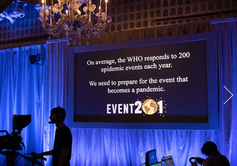

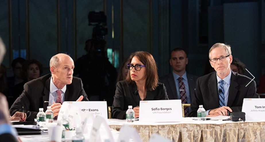

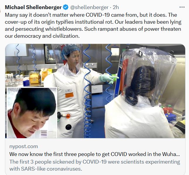


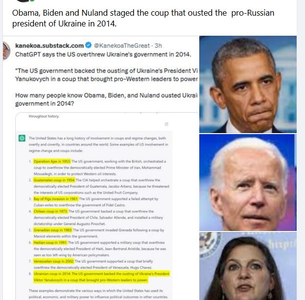
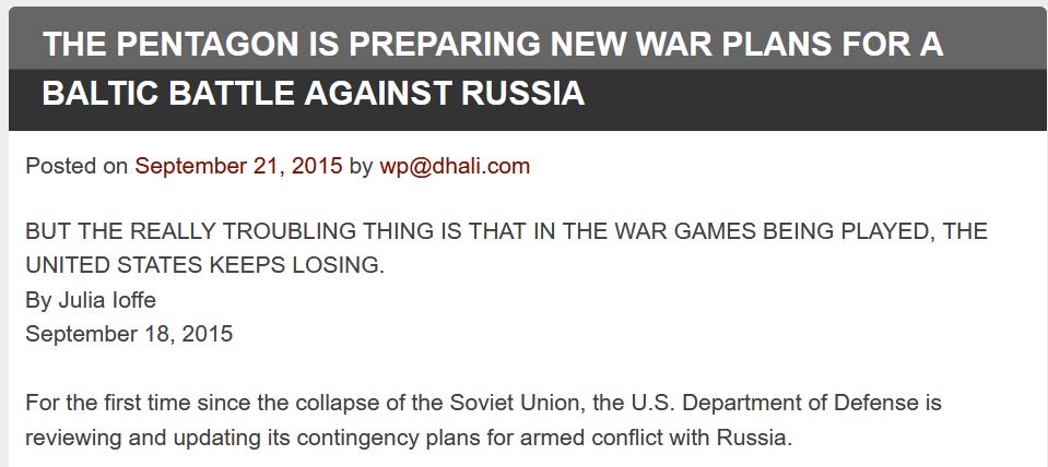
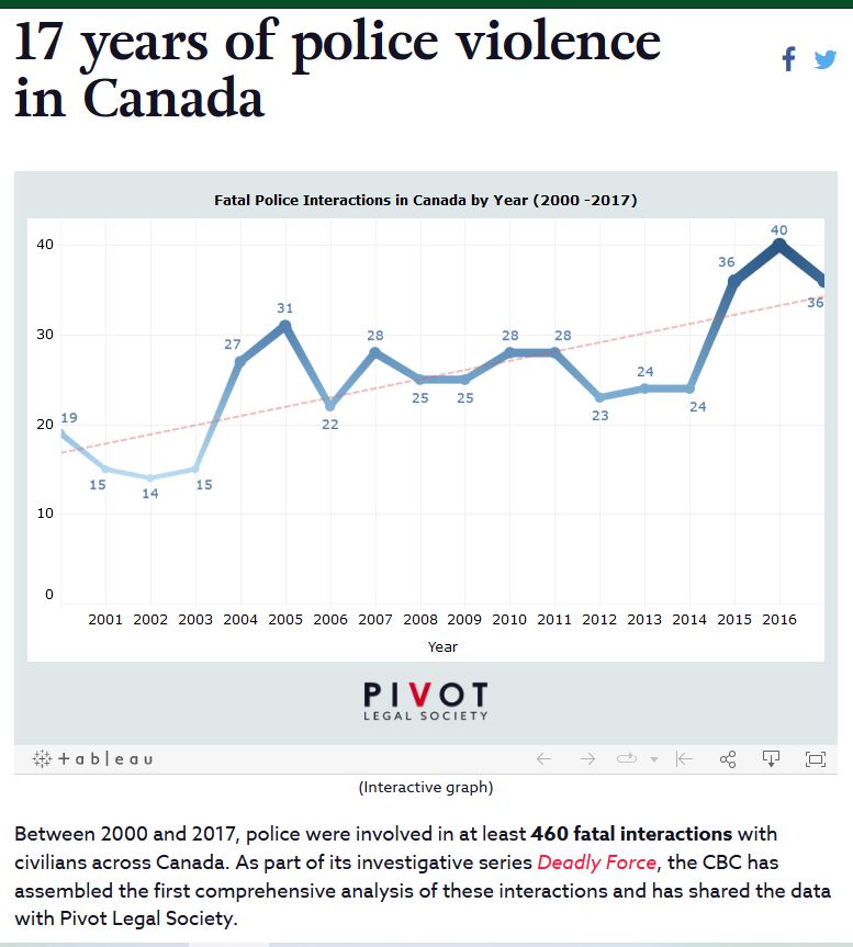

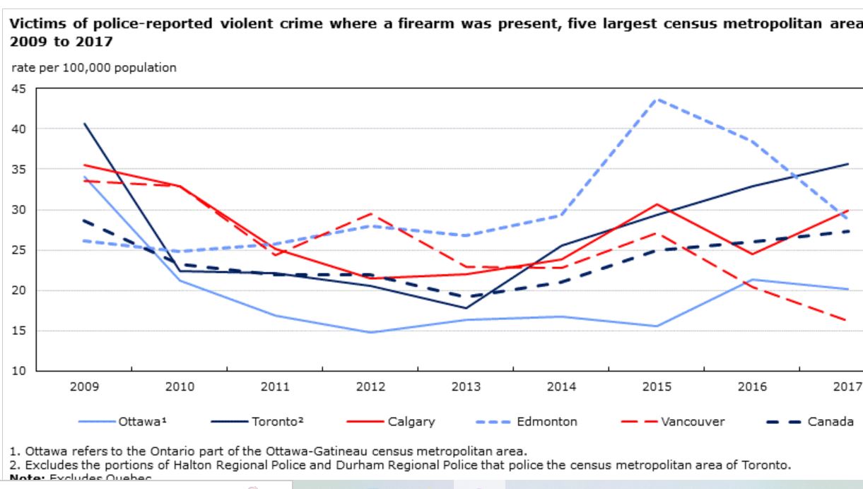


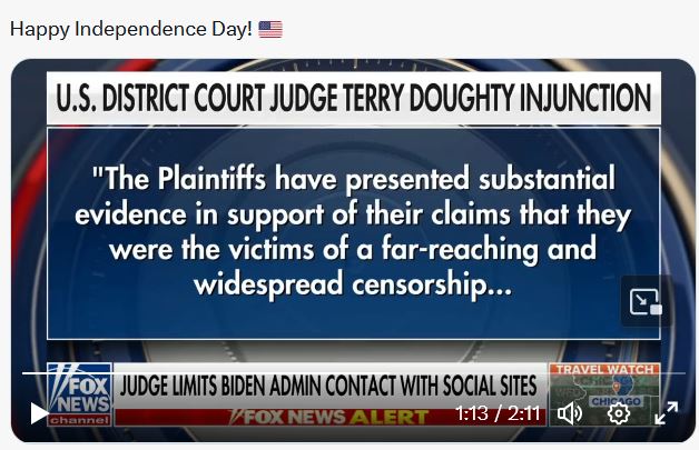


